- Call : +91 9540210956
- Email: [email protected]
- Artemis Hospital Sector 51 Gurgaon
- 09Am - 5Pm


Lung Function Tests Before Lung Surgery: FEV1, DLCO and Fitness Explained
- Home
- Blog Details
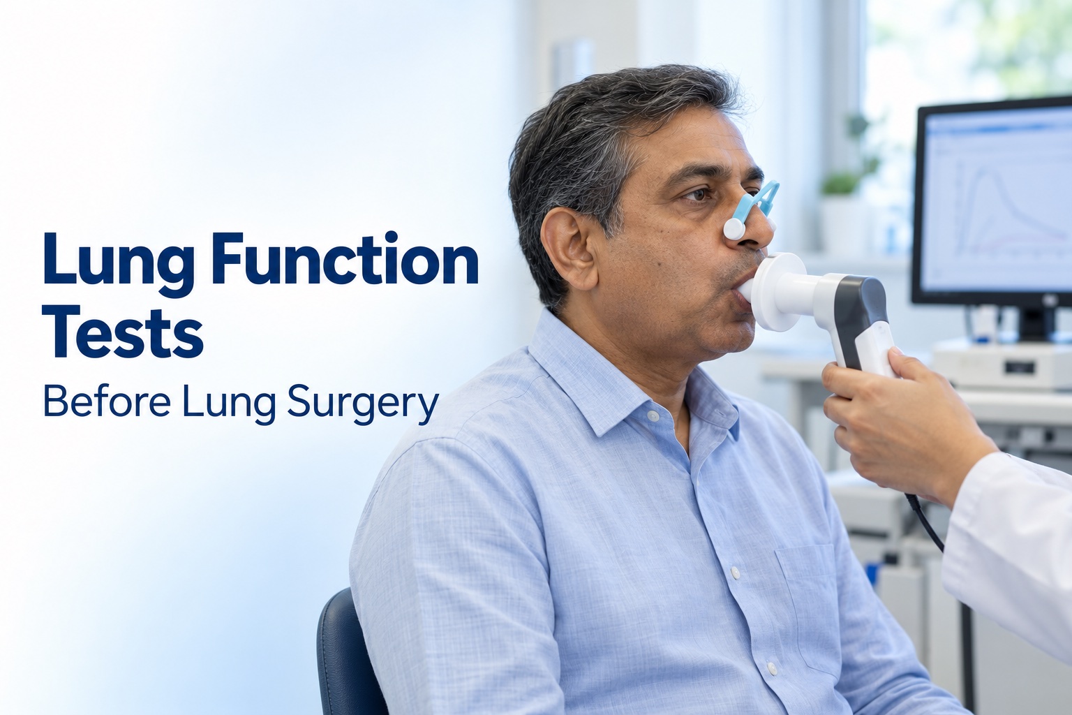
Before lung surgery, the team needs to answer two separate questions: can the proposed operation treat the disease, and can your body tolerate the operation and the lung function that remains afterwards? A scan helps with the first question. Breathing tests and a wider fitness assessment help with the second.
FEV1 measures airflow, DLCO measures gas transfer, and predicted postoperative values estimate reserve after the proposed resection. Exercise testing adds information about how your heart, lungs and circulation respond together under load. None of these measurements should become a standalone “pass” or “fail” label.
This guide explains the measurements, gives a worked example and separates current surgical risk categories from the interpretation of an ordinary lung function report. It draws on professional guidance, including the 2025 ERS/ESTS guideline, and published research on prediction and preparation.
Why Being Comfortable at Rest Is Not the Whole Assessment
Sitting comfortably or managing familiar daily activities does not show every aspect of lung reserve. Removing functioning lung tissue changes the demands on the remaining lung, and the extent of the proposed resection matters.
NICE lung cancer management guidance recommends spirometry and transfer-factor testing before curative-intent treatment, prediction of postoperative function before surgery, and additional exercise assessment where appropriate. The assessment is meant to inform treatment choices and discussion of breathlessness and complications.
A useful appointment should connect the measurements to your proposed operation. “Your breathing test is low” is incomplete: ask which measurement is low, what it means after the intended resection, and what additional information would help.
What Is Measured During Spirometry?
You take a full breath and then blow through a mouthpiece as forcefully and completely as instructed. The physiologist repeats the manoeuvre to check that the results are reproducible. Sometimes measurements are repeated after a bronchodilator.
| Measurement | Meaning | Why the distinction matters |
|---|---|---|
| FEV1 | Volume exhaled during the first second of a forced breath | Describes how quickly air can leave the lungs |
| FVC | Total volume exhaled during that forced manoeuvre | Provides context for the FEV1 result |
| FEV1/FVC | The proportion of the forced breath exhaled in the first second | Helps identify an airflow obstruction pattern |
The NHLBI pulmonary function laboratory guide describes spirometry and related testing. Good technique matters: tell the physiologist if you do not understand the instructions or need a break.
Does “70% Predicted” Mean Only 70% of My Lung Is Working?
No. “Percent predicted” compares a measurement with a reference value; it is not a count of how much lung tissue remains alive. Reference values account for characteristics such as age and height. Your report may also show a lower limit of normal or a z-score.
American Thoracic Society patient information explains the use of predicted values. A result expressed in litres and a result expressed as percent predicted are two ways of presenting a measurement, not two separate organs or functions.
Clinical interpretation also examines test quality and the pattern of results. The ERS/ATS technical standard on interpretation addresses reference limits and physiological patterns. The thresholds used to describe lung impairment are not interchangeable with the thresholds used to stratify surgical risk.
DLCO or TLCO: Why Airflow Alone Is Not Enough
The lungs must move air and transfer gas between the air sacs and bloodstream. DLCO, also called TLCO, measures transfer using a very small amount of carbon monoxide in a controlled test-gas mixture. It is a proxy for gas transfer, not a direct measurement of oxygen saturation.
A person can have an acceptable airflow result and still have impaired gas transfer. Conversely, the pattern of airflow and transfer results may differ between diseases. This is why one test does not substitute for the other.
During the test, you inhale through a mouthpiece, briefly hold your breath, and exhale as instructed. Factors such as anaemia and existing lung disease may affect interpretation. Ask whether the result has been adjusted for haemoglobin when relevant. Cleveland Clinic's diffusion-testing guide explains these factors and the test experience.
What Does “Predicted Postoperative” Mean?
The abbreviation ppo means predicted postoperative. Your team estimates the airflow and gas transfer expected after the planned resection, often using the contribution of the functioning lung to be removed.
One anatomical approach uses the proportion of functioning segments expected to remain. More detailed functional imaging may be useful when function is unevenly distributed. Published research comparing segment-based, scintigraphic and quantitative CT methods illustrates why different prediction methods exist.
Worked example, for explanation only: suppose an FEV1 is 80% predicted, all 19 segments are assumed to contribute equally, and three functioning segments are to be removed. A simplified estimate is 80 × 16/19, or approximately 67% predicted. This does not mean the patient will lose 13% of their ability to walk, and it does not give their complication probability.
That calculation depends on assumptions. An obstructed or severely damaged area may contribute differently from healthy tissue. Your team should explain whether a simple segment calculation is suitable for your anatomy and planned operation.
How Current Guidelines Group Predicted Results
The 2025 ERS/ESTS clinical practice guideline suggests the following categories for both ppoFEV1 and ppoDLCO:
| Predicted postoperative value | Guideline risk category |
|---|---|
| 60% predicted or higher | Normal risk category |
| 40–59% predicted | Moderate risk category |
| Below 40% predicted | High risk category |
These recommendations are conditional and based on very low-certainty evidence. “Normal risk category” does not mean no surgical risk. A high-risk result prompts further assessment and a discussion of alternatives; it is not, by itself, a universal prohibition on treatment.
The guideline considers wider health factors and patient preferences too. If your ppoFEV1 and ppoDLCO fall in different categories, ask the team to discuss both rather than relying on the more reassuring result.
What Extra Information Does Exercise Testing Provide?
A supervised walking test can assess functional capacity beyond resting measurements. NICE describes shuttle walking and cardiopulmonary exercise testing as options when further assessment of fitness is needed.
CPET commonly involves cycling on a stationary bicycle while resistance gradually increases. A fitted mask measures breathing gases, and staff monitor the ECG, blood pressure and oxygen level. The team observes how the cardiovascular and respiratory systems respond as activity becomes more demanding.
Cambridge University Hospitals' CPET guide explains the procedure and monitoring. The full appointment is longer than the exercise period because it includes preparation and recovery.
The resulting report contains several measurements; peak oxygen uptake is one of them. Ask what limits your exercise performance and how the findings change the proposed plan. Do not use an unsupervised stair-climbing challenge as a substitute for a test ordered by your team.
Why Predictions Are Not Recovery Guarantees
A predicted value estimates function under specified assumptions and at a particular stage of recovery. It cannot precisely tell you when you will walk a certain distance, return to work or stop feeling breathless.
A 2024 study of anatomical prediction after minimally invasive resections reported that formula-based estimates could overstate the eventual loss of FEV1 and DLCO in its study population. This is evidence of prediction limits, not proof that every patient will outperform their estimate.
Ask separately about short-term surgical risk and longer-term function. Your personal goals should be part of that conversation: carrying shopping, using stairs and returning to physically demanding employment are different recovery targets.
Can Fitness Be Improved Before Surgery?
Preparation may include a tailored exercise programme, smoking cessation support and review of existing conditions. The aim is to address modifiable problems within the treatment timetable, rather than postpone cancer treatment indefinitely to pursue a target number.
A 2022 Cochrane review included 10 trials with 636 participants. Preoperative exercise reduced postoperative pulmonary complications compared with no preoperative exercise: risk ratio 0.45, with a 95% confidence interval of 0.33–0.61 in nine trials. This is a relative reduction, not a 55-percentage-point reduction in an individual's risk.
The review also found a likely reduction in hospital stay, but results varied and evidence was uncertain for some outcomes. Ask for a supervised or prescribed programme suited to your symptoms and time before treatment. Do not assume that hard training on your own reproduces the studied interventions.
How to Prepare for the Tests
- Read the instructions for the specific tests booked; spirometry and CPET may have different medicine instructions.
- Bring your inhalers and a list of prescribed and non-prescribed medicines.
- Check whether an inhaler needs to be withheld and for how long; do not stop it simply because you are having a breathing test.
- Follow the laboratory's advice about smoking, meals, alcohol and exercise before testing.
- Wear loose clothing and suitable shoes if exercise testing is planned.
- Tell the team about recent infection, surgery, a heart event, chest pain or coughing up blood before starting.
NHS spirometry guidance explains why staff check recent health problems and whether it is appropriate to proceed. Tell them immediately if you feel faint, develop pain or cannot continue comfortably.
How Results May Change the Treatment Discussion
The review may lead to another test, optimisation of an existing condition, or a more detailed comparison of treatment choices. Where surgery remains an option, ask which extent of resection is being considered and how that affects the prediction.
Our segmentectomy versus lobectomy guide explains the operation choices. A smaller resection should not be selected from a breathing-test number alone: the proposed cancer treatment still needs to be appropriate.
Ask for the reasoning in concrete terms: “What would you recommend if the exercise test is reassuring, and what would change if it is not?” A clear answer is more useful than a percentage presented without a decision attached to it.
Questions to Take to Your Appointment
- What are my FEV1 and DLCO, and were the tests technically reliable?
- What are my predicted postoperative values for the planned operation?
- Which assumptions were used in the calculation?
- Do I need functional imaging or an exercise test?
- Which risks come from my lungs, and which come from other health conditions?
- Would a preparation programme help within my treatment timetable?
- What are the alternatives if the proposed operation carries too much risk?
- What breathing limitations should I realistically plan for afterwards?
Frequently Asked Questions
Does a low FEV1 automatically rule out surgery?
No. Ask for a combined assessment of airflow, gas transfer, predicted remaining function, exercise capacity and the rest of your health.
Can normal oxygen saturation replace DLCO?
No. A resting oxygen reading and a diffusion test measure different aspects of breathing. Ask why each test has been requested in your case.
Is 60% a universal pass mark?
No. The table describes guideline risk categories, not a guarantee or a rule that applies independently of all other findings.
Will breathing exercises make my result normal?
Do not expect a guaranteed change in a measurement. Ask what your prescribed exercises are intended to improve and how progress will be assessed.
Arranging a Surgical Review
Bring the complete lung function report, recent scans and treatment history. Our preparation checklist can help organise these. For a thoracic surgical opinion, book a consultation with Dr. Parveen Yadav to discuss how your findings affect the proposed operation.
Selected Research and Guidelines
- ERS/ESTS clinical practice guideline, 2025: current fitness assessment and predicted postoperative risk categories; recommendations include evidence-certainty qualifications.
- NICE NG122 management guidance: lung function, postoperative prediction and further fitness assessment.
- ERS/ATS technical standard, 2022: interpretation of routine lung function tests.
- Granger and Cavalheri, Cochrane, 2022: benefits and limitations of preoperative exercise evidence.

Dr. Parveen Yadav
18+ Yrs Exp | 5,700+ Thoracic & Robotic Cancer Surgeries
Dr. Parveen Yadav is a Director and Senior Consultant in Thoracic and Surgical Oncology, specializing in minimally invasive and robotic lung and esophageal surgeries, with advanced training from AIIMS and Tata Memorial Hospital.
View Full ProfileRecent Posts
Does a Positive PET-CT Always Mean Lung Cancer?
- Dr. Parveen Yadav
- Oct 14, 2026
Chest Drain After Lung Surgery: Care, Removal and Recovery
- Dr. Parveen Yadav
- Oct 10, 2026
EBUS Biopsy: What to Expect and How It Helps Stage Lung Cancer
- Dr. Parveen Yadav
- Oct 06, 2026
Rib Pain When Coughing: Causes, Warning Signs and Treatment
- Dr. Parveen Yadav
- Oct 02, 2026
Related Posts
Chest Drain After Lung Surgery: Care, Removal and Recovery
Understand why a chest drain is used after lung surgery, how to move safely with it, what guides removal and which warning signs need urgent help.
- Read More
- Oct, 10 2026
Recurrent Pneumothorax: When Are VATS and Pleurodesis Needed?
Learn when recurrent pneumothorax needs surgery, how VATS and pleurodesis help prevent another collapsed lung, and what recovery and follow-up involve.
- Read More
- Sep, 24 2026
Fluid Around the Lung (Pleural Effusion): Causes and Treatment
Understand pleural effusion (fluid around the lung): what causes it, common symptoms, how it is investigated and why treatment depends on the underlying cause.
- Read More
- Sep, 17 2026
Copyright 2026 © Dr .Parveen Yadav all rights reserved.
Proudly Scaled by Public Media Solution!