


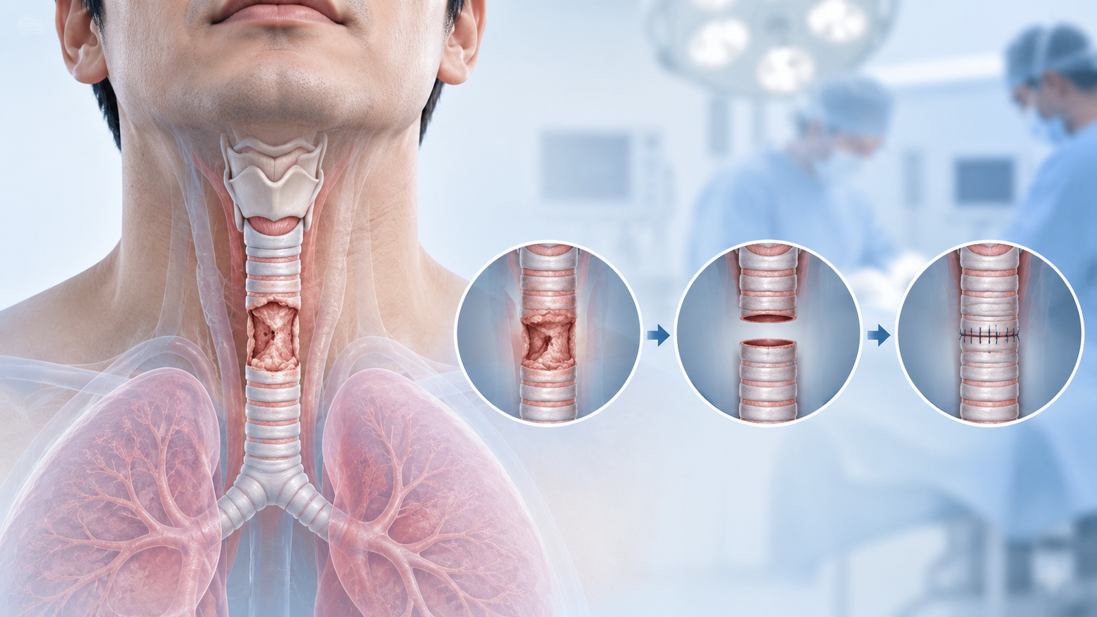
Removing a damaged segment of the windpipe and rejoining the healthy ends sounds more alarming than it usually is in practice. For the right patient, tracheal resection and reconstruction is a well-established operation with a high success rate, and understanding the steps involved makes the decision to go ahead far less daunting.
Tracheal resection and reconstruction is a surgery in which a diseased or narrowed segment of the trachea, the windpipe carrying air from the throat to the lungs, is removed, and the two healthy ends are then rejoined directly. It is used when a section of the airway has been damaged badly enough that stretching, stenting, or repeated endoscopic procedures are no longer a durable solution.
The trachea has some natural give, allowing the neck and chin to be positioned in a way that takes tension off the join while it heals, which is why segments of a reasonable length can usually be removed and rejoined without needing an artificial graft.
Not every airway narrowing needs resection. This surgery is generally considered when the problem is localised, meaning it affects a defined segment rather than the whole trachea, and other options have already been tried or are unlikely to work well.
The approach depends on where the narrowed or diseased segment sits. A stenosis in the neck region is usually approached through a collar incision at the base of the neck. A segment lower down, closer to where the trachea branches into the lungs, may need an approach through the chest, sometimes combined with the neck incision for a longer segment.
Once the diseased segment is identified, it is carefully removed, and the two healthy ends of the trachea are sewn back together, an anastomosis, using fine sutures placed with meticulous technique, since this join is the single most critical part of the operation.
To reduce tension on the fresh join while it heals, the surgical team commonly places a temporary stitch from the chin to the chest, keeping the neck gently flexed forward for the first several days after surgery. This sounds uncomfortable, and it is described that way by most patients, but it is a short-term measure that protects a much more important long-term outcome.
Most patients spend the first day or two in a high-dependency or intensive care setting for close airway monitoring, since the early period after surgery is when careful observation matters most.
The chin-to-chest stitch, where used, typically stays in place for about a week, and patients are specifically counselled to avoid extending the neck backward during this time. Something as simple as looking up sharply is avoided.
Voice changes and mild difficulty swallowing are common in the initial days, usually improving steadily over one to two weeks as swelling around the airway settles.
Most patients are eating a normal diet and speaking clearly well before discharge, and hospital stays are typically in the range of a week, depending on the extent of surgery and how smoothly the initial recovery goes.
A follow-up bronchoscopy, a look inside the airway with a thin camera, is usually done some weeks after surgery to directly check how the join has healed.
Overall, roughly 95 percent of patients who undergo tracheal resection and reconstruction have a good result, which makes this one of the more reliably successful major airway operations. That said, it is major surgery, and being clear-eyed about the risks is part of making an informed decision.
Complications occur in around one in five patients, and about half of those relate specifically to the join itself, ranging from minor, self-resolving issues to, less commonly, a leak or narrowing at the anastomosis that needs further treatment.
Other recognised risks include infection, bleeding, temporary or, rarely, lasting voice changes from nerve irritation near the airway, and swelling that can temporarily affect breathing in the immediate post-operative period.
Certain factors are known to raise the risk of a complication at the join:
This is exactly why a careful pre-operative work-up, mapping the exact length and location of the narrowed segment, matters so much before deciding on surgery, and why this type of operation is best done in a centre with specific experience in airway surgery.
For most patients, the impact of a successful tracheal resection and reconstruction is substantial: breathing returns to normal without the noisy, effortful quality that often comes with airway narrowing, and the daily limitations that come with a partially blocked airway lift considerably.
Where voice quality was affected by the obstruction itself, this often improves too, once the airway is fully open again.
Follow-up continues for some time afterward, both to confirm the join is healing well and to watch for any early signs of recurrent narrowing, which is uncommon but is easier to manage early if it does occur.
If you have been told you have tracheal stenosis, a tracheal tumor, or have had repeated procedures for a narrowed airway that keep needing to be repeated, it is worth a dedicated consultation to discuss whether resection and reconstruction is the right next step for your specific situation, rather than continuing with temporary measures.
Dr. Parveen Yadav is a thoracic surgeon based at Artemis Hospital, Sector 51, Gurgaon, with experience in complex airway, lung, and esophageal surgery.
If you are dealing with a tracheal narrowing or tumor and want a clear opinion on your options, book a consultation to review your scans and airway history together.

18+ Yrs Exp | 5,700+ Thoracic & Robotic Cancer Surgeries
Dr. Parveen Yadav is a Director and Senior Consultant in Thoracic and Surgical Oncology, specializing in minimally invasive and robotic lung and esophageal surgeries, with advanced training from AIIMS and Tata Memorial Hospital.
View Full Profile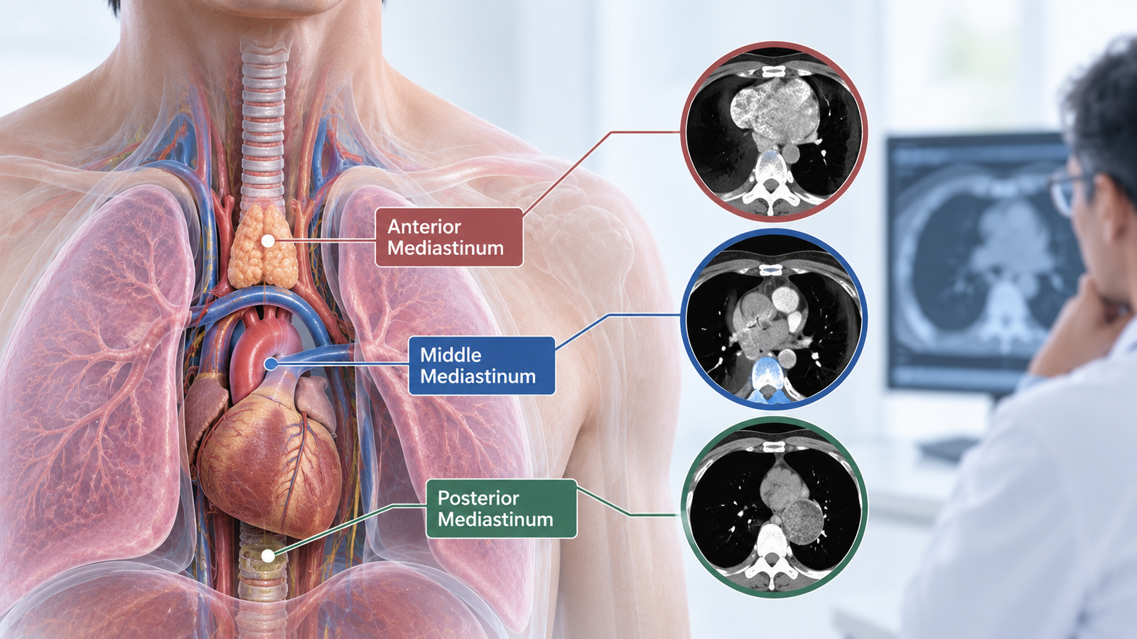
Learn how the location of a mediastinal mass in the anterior, middle, or posterior mediastinum helps doctors narrow the diagnosis and plan further tests or biopsy.
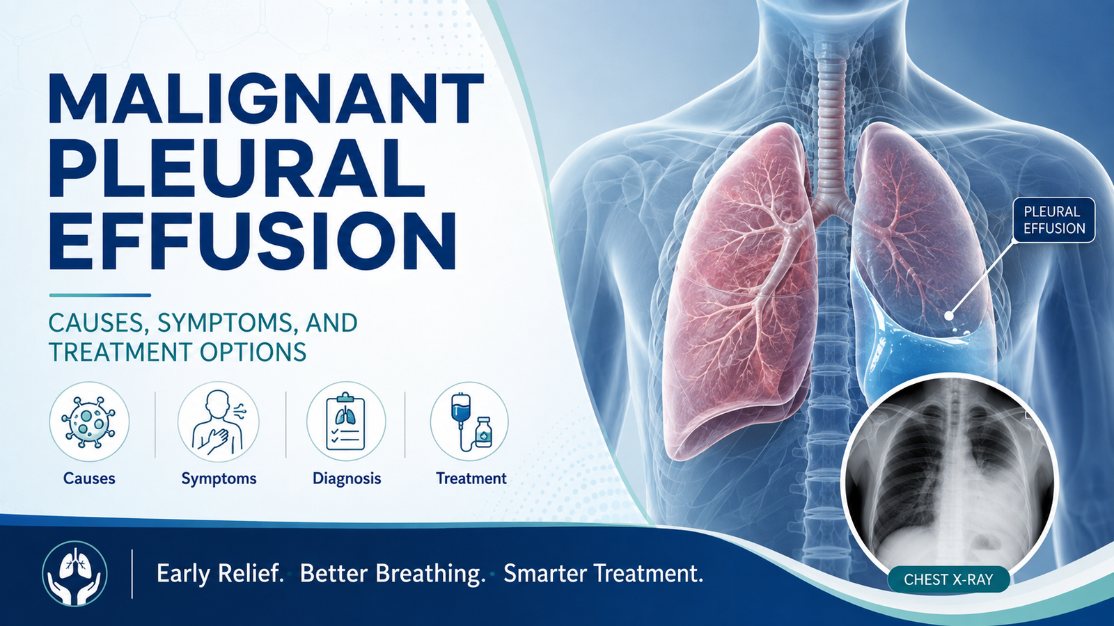
Learn what causes malignant pleural effusion in lung cancer, its symptoms, and treatment options including thoracentesis, pleurodesis, and PleurX catheters.
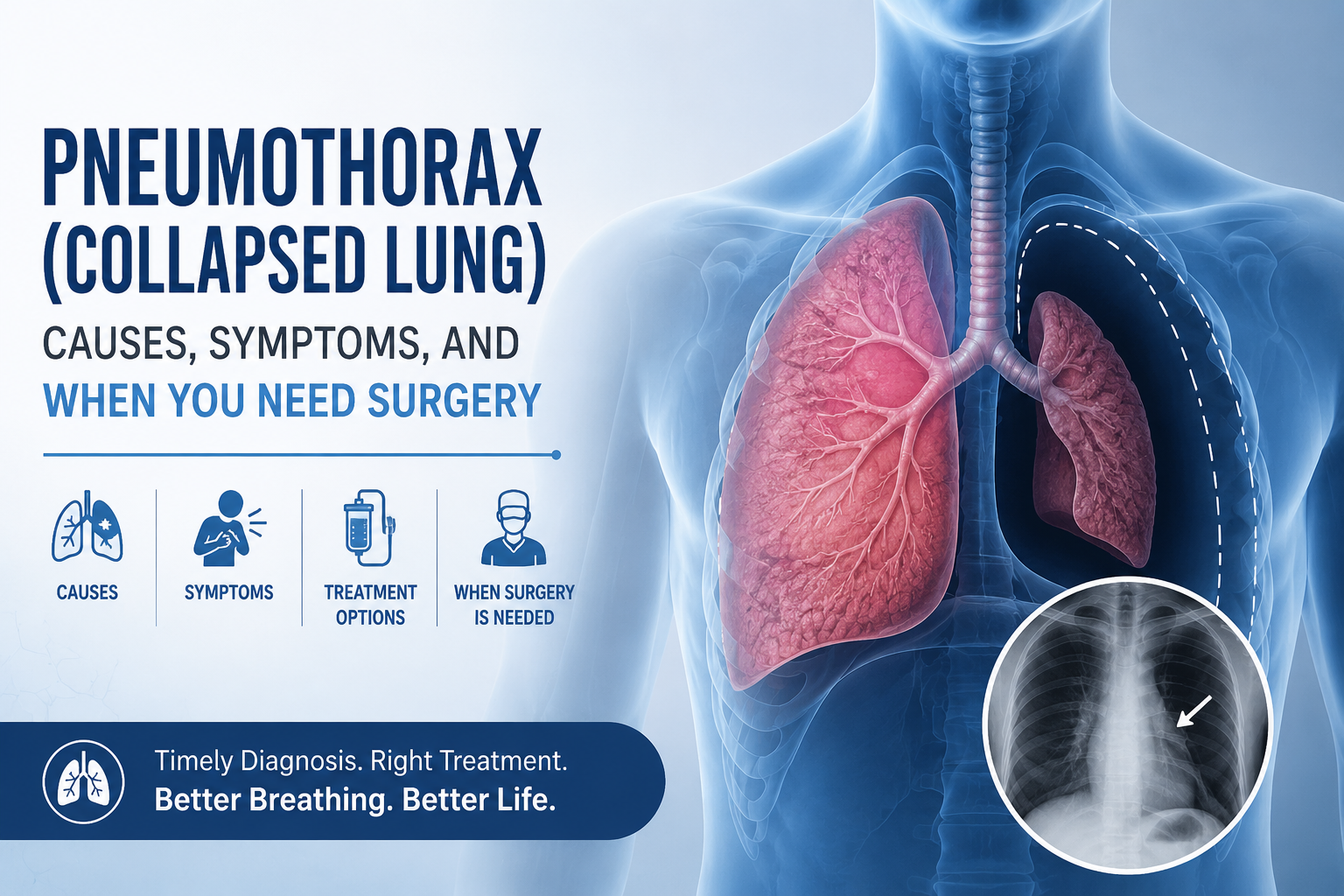
Learn pneumothorax (collapsed lung) causes, symptoms, chest tube treatment, and when VATS surgery is needed to prevent it from coming back.
Copyright 2026 © Dr .Parveen Yadav all rights reserved.
Proudly Scaled by Public Media Solution!