


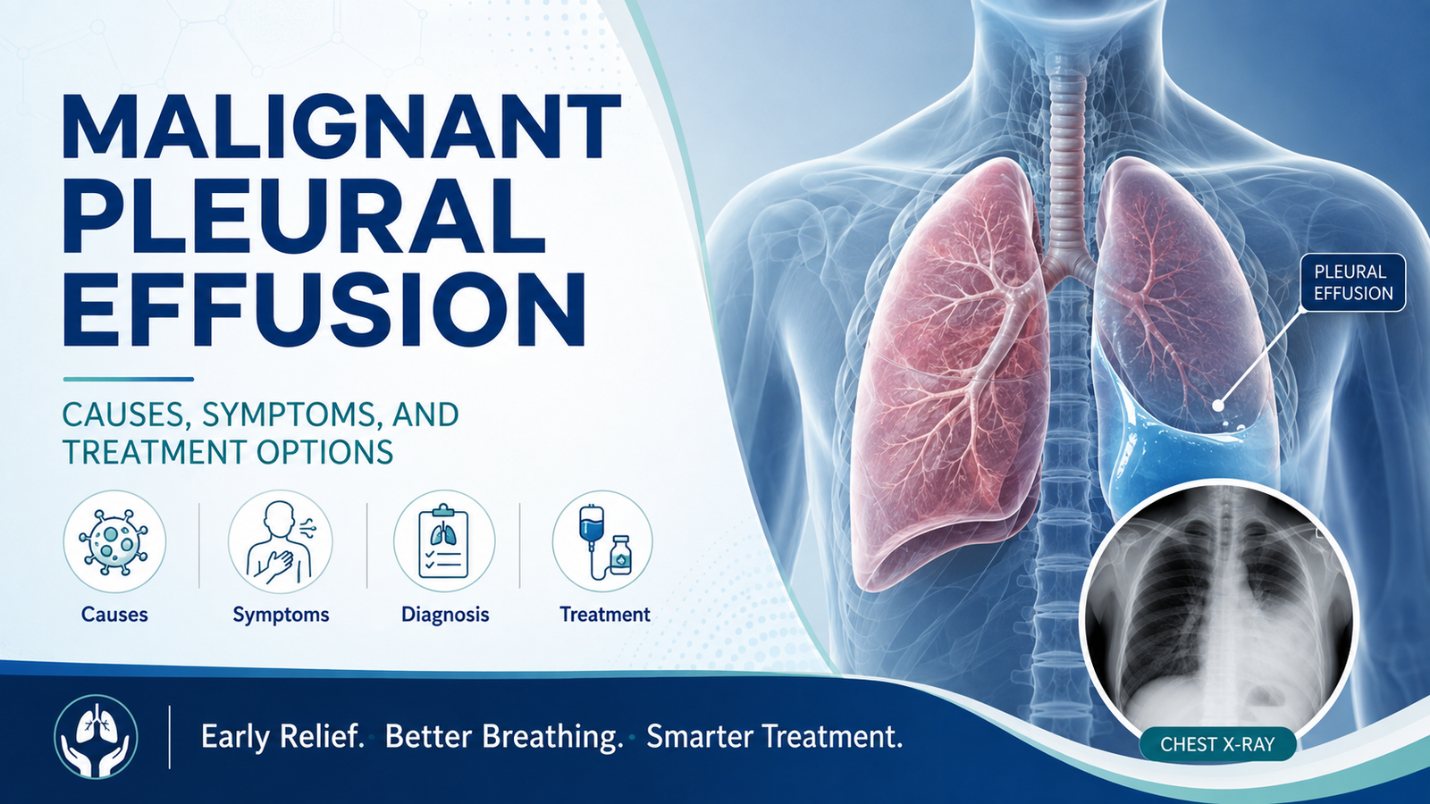
A malignant pleural effusion is a build-up of fluid around the lung caused directly by cancer, most often lung cancer, breast cancer, or cancer that has spread from another organ. It is one of the most common reasons a cancer patient suddenly develops breathlessness, and the fluid usually keeps coming back unless it is specifically treated, not just drained once. The right treatment depends less on how much fluid is present and more on how well the lung re-expands once the fluid is removed, and what the overall cancer treatment plan looks like.
This guide explains why malignant pleural effusion happens, how it is diagnosed, and the treatment options doctors choose between, including thoracentesis, talc pleurodesis, and indwelling pleural catheters such as PleurX.
The pleura is the thin lining around the lung and inside the chest wall. Normally it holds only a small amount of fluid that helps the lung glide smoothly during breathing. A pleural effusion develops when fluid builds up beyond this small amount.
A pleural effusion is called malignant when cancer cells are found directly in the fluid or on the pleural lining itself, meaning the cancer has spread to the pleura. This is different from a pleural effusion caused indirectly by a tumour blocking lymphatic drainage or by pneumonia, heart failure, or low protein levels, which are managed very differently.
Almost any cancer can spread to the pleura, but a small number of cancers account for most cases.
Symptoms depend on how quickly the fluid has built up and how much is present, since a slowly accumulating effusion can sometimes reach a large volume before symptoms become severe.
Because breathlessness in a cancer patient can also come from anaemia, cancer-related fatigue, lung involvement by the cancer itself, or unrelated heart or lung disease, imaging is needed to confirm that a pleural effusion is the specific cause before planning drainage.
There is no single "best" treatment for every patient. The choice depends on how breathless the patient is, whether the lung fully re-expands after drainage, how much fluid reaccumulates, expected survival, and whether ongoing cancer treatment (chemotherapy, targeted therapy, or immunotherapy) is likely to control the fluid on its own.
Lung re-expansion after drainage: Talc pleurodesis is favoured when the lung re-expands fully. PleurX catheter is favoured when the lung does not fully re-expand ("trapped lung").
Expected fluid reaccumulation: Slower reaccumulation favours talc pleurodesis, whereas fast, frequent reaccumulation favours PleurX.
Ability to tolerate a hospital stay: Talc pleurodesis suits patients fit for a short admission; PleurX is preferred for patients who wish to avoid a hospital stay or have limited mobility.
Access to home drainage support: PleurX requires a caregiver available to help with home drainage.
Neither option is universally "better." A patient whose lung does not re-expand after drainage (a trapped lung) will not benefit from pleurodesis, since the two pleural surfaces cannot properly fuse together, making an indwelling catheter the more logical choice in that specific situation.
In India, chest tube-based talc pleurodesis remains the more widely available and commonly used option in most centres, partly because indwelling pleural catheters are imported devices and can be significantly more expensive, with less insurance coverage and fewer trained home-care nursing services to support long-term outpatient drainage compared to some Western healthcare systems.
VATS-guided talc poudrage is increasingly available at centres with thoracic surgery expertise and offers a practical, cost-effective alternative that combines direct visualisation, biopsy capability, and a high pleurodesis success rate in a single admission.
For patients who specifically need an indwelling catheter, for example those with a trapped lung, it is worth discussing availability and cost directly with your treating team early rather than after multiple failed drainage attempts.
Malignant pleural effusion is a common and very treatable complication of advanced cancer, even when it cannot be cured outright, because relieving breathlessness meaningfully improves day-to-day quality of life. The right approach, whether that is talc pleurodesis, VATS with talc poudrage, or an indwelling pleural catheter, depends on how the lung behaves after drainage, practical factors like caregiver support, and where you are in your overall cancer treatment.
If you or a family member has developed breathlessness with a known or suspected pleural effusion, book a consultation with Dr. Parveen Yadav to review your scans and fluid reports and discuss which drainage strategy fits your specific situation. Bring your chest X-ray or CT scan and any prior fluid cytology reports to your appointment so the right approach can be planned without delay.
Related reading:
For advanced lung cancer treatment options, see our Lung treatment page.

18+ Yrs Exp | 5,700+ Thoracic & Robotic Cancer Surgeries
Dr. Parveen Yadav is a Director and Senior Consultant in Thoracic and Surgical Oncology, specializing in minimally invasive and robotic lung and esophageal surgeries, with advanced training from AIIMS and Tata Memorial Hospital.
View Full Profile Pneumothorax (Collapsed Lung): Causes, Symptoms, and When You Need Surgery
 Surgery vs. Endoscopic Resection for T2N0M0 Lung Cancer: What’s the Safer Option?
 Robotic Lobectomy vs. Traditional VATS: An Expert’s Guide
Learn pneumothorax (collapsed lung) causes, symptoms, chest tube treatment, and when VATS surgery is needed to prevent it from coming back.
T2N0M0 lung cancer diagnosis? Compare surgery vs. non-surgical options like SBRT. Learn about safety, survival rates, and what's the safer choice for you.
Robotic vs VATS lobectomy? An expert guide to choosing the best lung cancer treatment in Gurgaon & Delhi. Learn the pros, cons, and key differences.
Copyright 2026 © Dr .Parveen Yadav all rights reserved.
Proudly Scaled by Public Media Solution!