


Two CT reports can both say "mediastinal mass" and mean almost entirely different things. Before any biopsy is even planned, the single most useful piece of information a thoracic surgeon looks at is where in the mediastinum the mass actually sits, because location alone narrows the likely diagnosis dramatically.
The mediastinum is the central space in the chest, sitting between the two lungs, and it houses the heart, major blood vessels, the trachea, the esophagus, the thymus gland, and numerous lymph nodes and nerves. Because so many different structures share this one space, a mass here could, in principle, arise from almost anything.
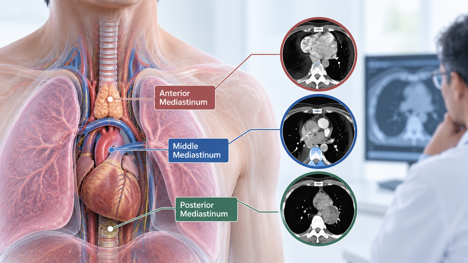
That is exactly why thoracic surgeons and radiologists split the mediastinum into compartments, anterior, middle, and posterior, based on which structures normally sit in each zone.
A mass's compartment does not confirm the diagnosis on its own, but it changes the list of likely possibilities enormously, and it is one of the first things noted on any chest CT report describing a mediastinal mass.
The anterior mediastinum sits between the back of the breastbone and the front of the pericardium, the sac around the heart. It normally contains the thymus gland, fatty tissue, and lymph nodes, and it is, by a clear margin, the most frequent site for a mediastinal mass overall, accounting for slightly more than half of all cases.
The two dominant possibilities in this compartment are thymic tumors, thymoma being the most common single primary tumor of the anterior mediastinum, and lymphoma.
Less commonly, germ cell tumors occur here, along with rarer possibilities such as an extension of an enlarged thyroid gland reaching down into the chest, or ectopic parathyroid tissue.
A mass in this compartment in a middle-aged adult raises thymoma fairly high on the list; in a younger patient, lymphoma or a germ cell tumor becomes relatively more likely, age and compartment together already doing significant diagnostic work before any tissue sample is taken.
The middle mediastinum sits between the anterior compartment and the front of the spine, and it is a genuinely crowded space, containing the heart and pericardium, the major vessels entering and leaving the heart, the trachea and main airways, and a large number of lymph nodes.
Around a quarter of all mediastinal masses are found here, and roughly two-thirds of those are cysts, most often foregut duplication cysts or pericardial cysts, both of which are benign and often simply monitored rather than removed unless they cause symptoms or grow.
Enlarged lymph nodes are the other common finding in this compartment, which can reflect infection, sarcoidosis, or, particularly if the pattern and imaging features raise concern, lymphoma or spread from a lung cancer.
The posterior mediastinum lies behind the heart and trachea, extending back toward the spine, and contains the esophagus, the descending aorta, and a dense collection of nerve tissue, including the sympathetic chain and nerve roots.
This changes the diagnostic picture substantially: roughly four out of five masses found here are neurogenic tumors, arising from nerve tissue, and these are frequently benign, particularly in adults.
In children, a neurogenic mass in this location raises a different and more specific set of considerations, which is one reason age is always read alongside location rather than location alone.
Knowing the likely compartment shapes almost every decision that follows. It influences which additional tests are ordered, whether a PET scan or tumor markers add useful information, how urgently a biopsy is pursued, and even how the biopsy itself is best performed, since some compartments are more safely and easily accessed through one approach than another.
It also shapes how a patient should reasonably feel about a report describing a mediastinal mass.
A cyst discovered incidentally in the middle mediastinum on a scan done for an unrelated reason carries a very different weight than a rapidly growing anterior mass in a young adult, even though both might appear as a similarly sized shadow on an initial chest X-ray.
Compartment, size, growth pattern on serial imaging, and the patient's age and symptoms are read together, not any single feature in isolation.
Many mediastinal masses, particularly smaller ones, cause no symptoms at all and are found incidentally on imaging done for another reason.
When symptoms do occur, they generally relate to a mass pressing on a nearby structure rather than to the mass itself, and the pattern can offer further clues.
Once a mass is found and its compartment identified, the typical next steps include a contrast CT scan for detailed anatomical mapping, sometimes an MRI when soft tissue detail near the spine or major vessels needs closer definition, and blood tests for tumor markers when a germ cell tumor is a realistic possibility based on compartment and age.
Tissue diagnosis, usually through a needle biopsy or, where appropriate, direct surgical removal that serves as both diagnosis and treatment, generally follows once imaging has narrowed the likely possibilities.
If a scan has identified a mediastinal mass, the most useful next step is a consultation that reviews the mass in the context of its exact location, your age, and your symptoms, rather than the finding in isolation.
Many mediastinal masses turn out to be benign, but the diagnostic pathway still needs to be followed properly to confirm that.
Dr. Parveen Yadav is a thoracic surgeon based at Artemis Hospital, Sector 51, Gurgaon, with specific experience in robotic and minimally invasive mediastinal surgery.
If you have a mediastinal mass on a recent scan, book a consultation for a clear, structured next-steps plan.

18+ Yrs Exp | 5,700+ Thoracic & Robotic Cancer Surgeries
Dr. Parveen Yadav is a Director and Senior Consultant in Thoracic and Surgical Oncology, specializing in minimally invasive and robotic lung and esophageal surgeries, with advanced training from AIIMS and Tata Memorial Hospital.
View Full Profile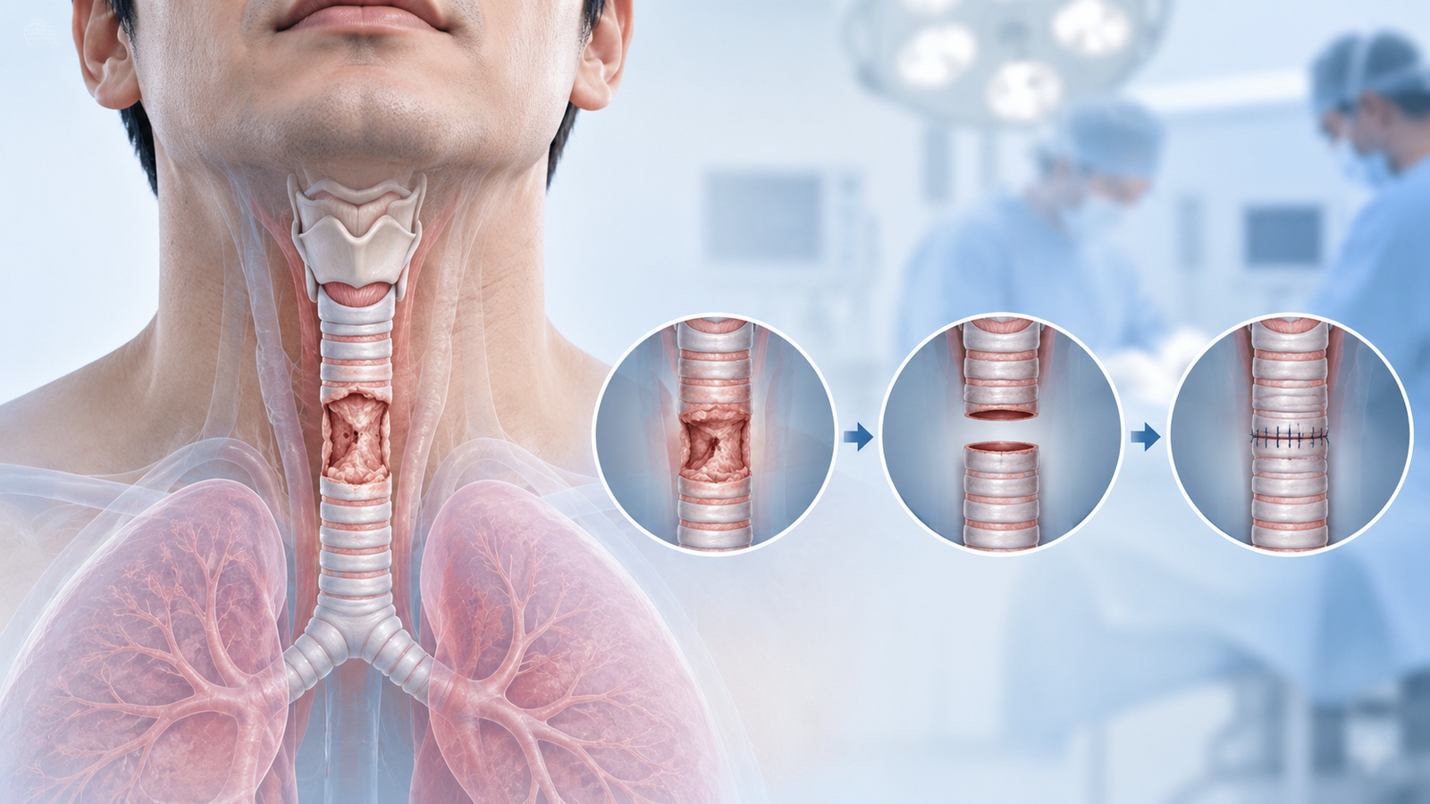 Tracheal Resection and Reconstruction: What Patients Should Know
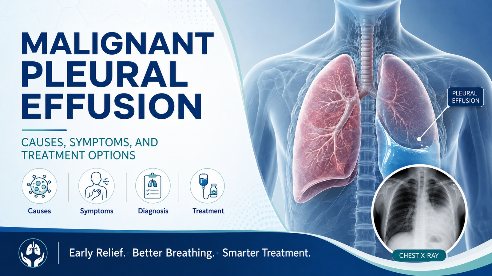 Malignant Pleural Effusion: Causes, Symptoms, and Treatment Options
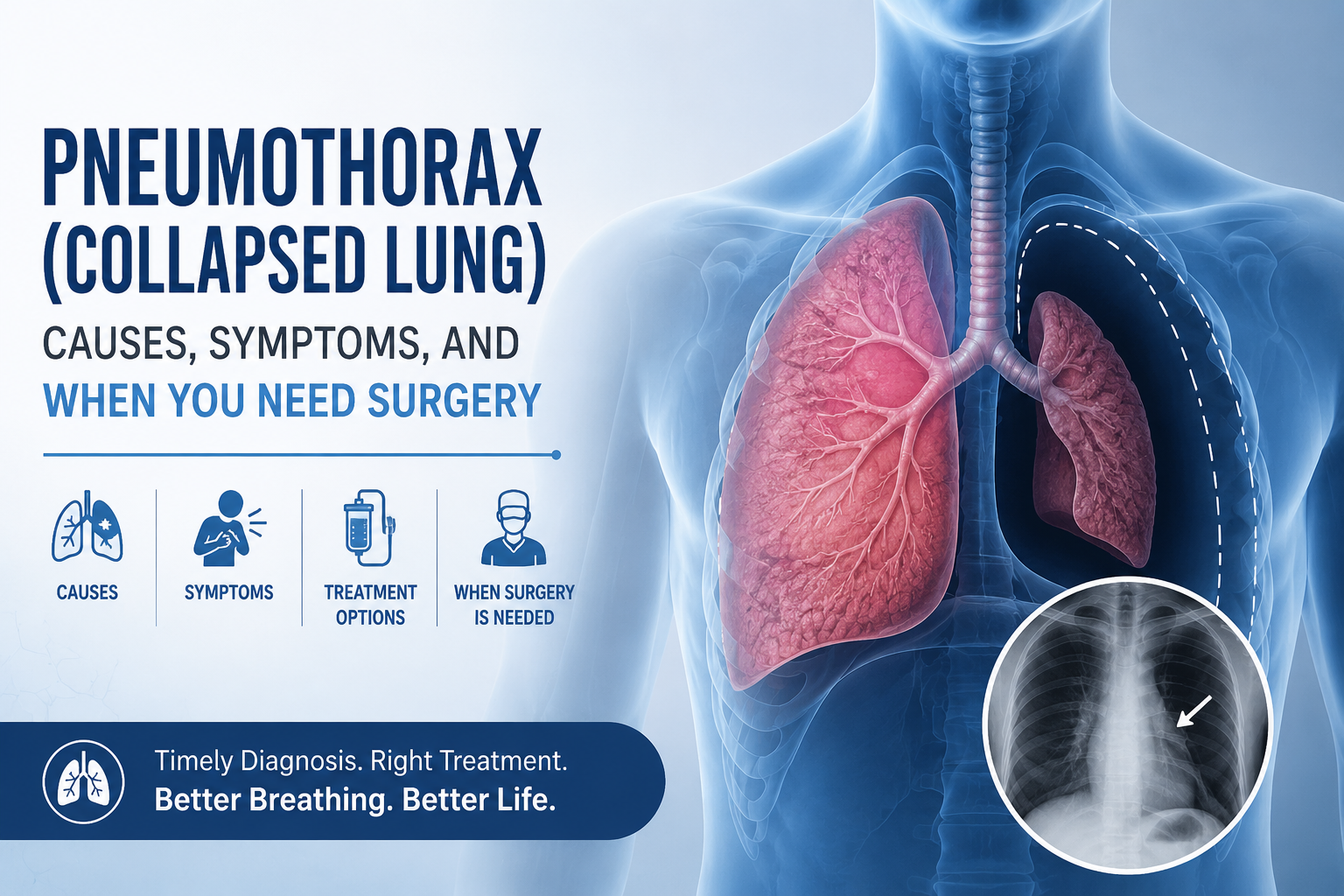 Pneumothorax (Collapsed Lung): Causes, Symptoms, and When You Need Surgery
Learn how tracheal resection and reconstruction is performed, who needs it, recovery time, risks, success rates, and when surgery is recommended for tracheal stenosis or tumors.
Learn what causes malignant pleural effusion in lung cancer, its symptoms, and treatment options including thoracentesis, pleurodesis, and PleurX catheters.
Learn pneumothorax (collapsed lung) causes, symptoms, chest tube treatment, and when VATS surgery is needed to prevent it from coming back.
Copyright 2026 © Dr .Parveen Yadav all rights reserved.
Proudly Scaled by Public Media Solution!