


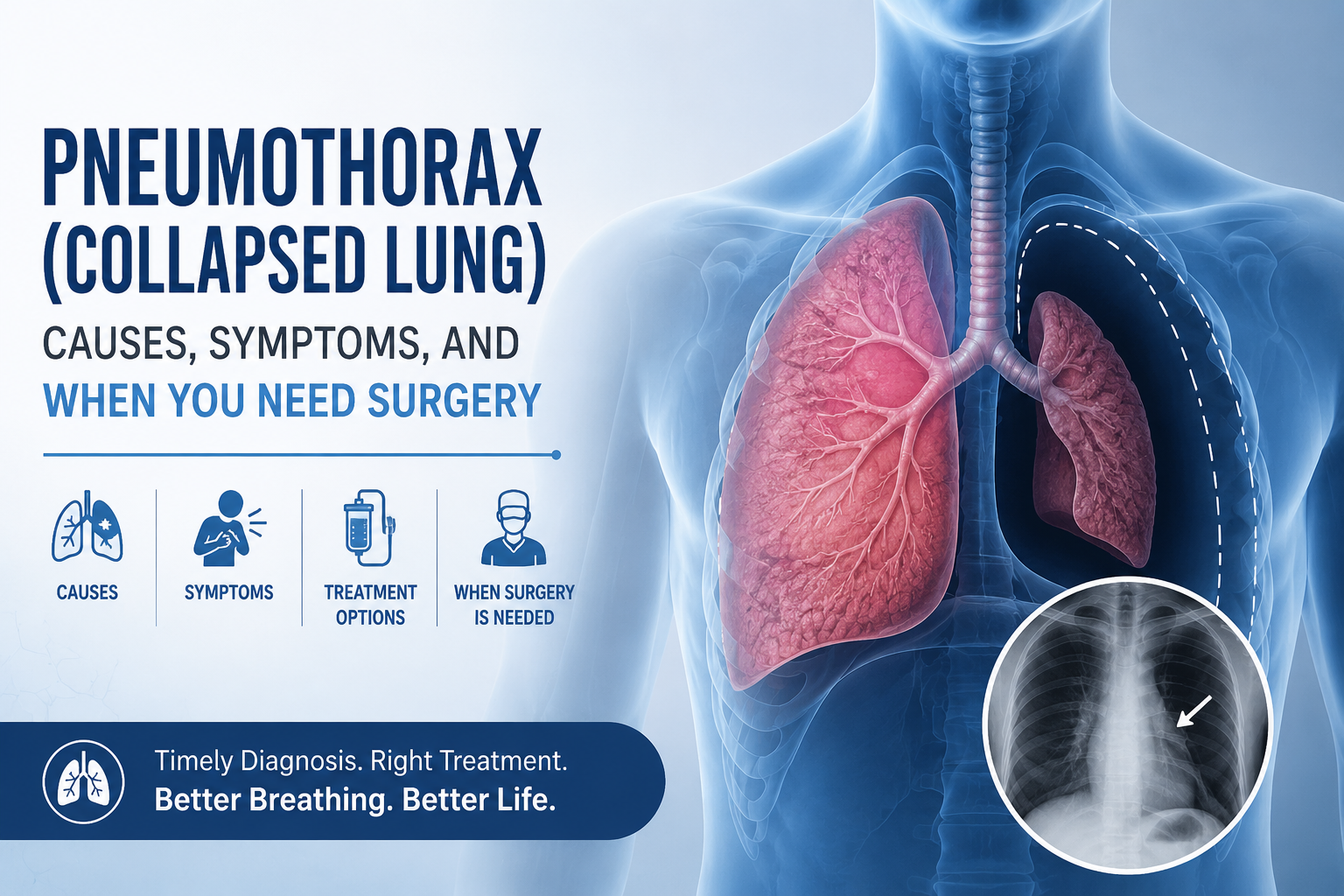
A pneumothorax, commonly called a collapsed lung, happens when air leaks into the space between the lung and the chest wall and pushes the lung inward instead of letting it expand. It can happen suddenly in a young, otherwise healthy person, after an injury, or as a complication of an existing lung disease such as COPD or old tuberculosis scarring. A first, small pneumothorax is often managed with observation or a chest tube, but a recurring or large one usually needs a procedure to stop it from happening again.
This guide explains what causes a pneumothorax, how doctors decide between watching, needle aspiration, a chest tube, or surgery, and why some patients in India need surgery earlier than textbook guidelines from Western countries suggest.
The lung sits inside the chest wall, separated from it by a thin, slippery layer called the pleura. Normally there is no air in this space, only a small amount of fluid that lets the lung slide smoothly as you breathe. A pneumothorax occurs when air enters this pleural space, either from a tear in the lung itself or from an injury through the chest wall. As air builds up, it presses on the lung from outside and causes part or all of it to collapse, making it harder to expand and take in oxygen.
Doctors broadly divide pneumothorax into a few categories, and the cause changes both the urgency and the long-term treatment plan.
Occurs in people with no known lung disease, most often tall, thin young men, usually caused by tiny air-filled blisters on the lung surface called blebs or bullae that rupture without warning.
Occurs in people with existing lung disease such as COPD, emphysema, or, very commonly in India, old healed tuberculosis that has left scarred, weakened lung tissue prone to rupture.
Caused by chest injury such as a road traffic accident, a fall, a stab or gunshot wound, or a fractured rib puncturing the lung.
A complication of a medical procedure such as a lung biopsy, central line placement, or ventilator support.
A rare but life-threatening emergency where trapped air keeps building pressure with no way to escape, pushing the heart and major blood vessels to one side and cutting off blood flow if not treated immediately.
In India, secondary spontaneous pneumothorax linked to old or active tuberculosis is far more common than in Western countries, which is one reason recurrence patterns and treatment decisions here often differ from Western textbook guidelines.
Symptoms usually start suddenly and vary depending on how much of the lung has collapsed.
Small pneumothoraxes can sometimes cause only mild discomfort and are found incidentally on a chest X-ray. A large or tension pneumothorax, on the other hand, can cause severe breathlessness and dangerously low oxygen levels within minutes and needs emergency treatment.
Call for emergency care immediately if there is severe or worsening breathlessness, bluish discoloration of lips or skin, chest pain that is severe or spreading, confusion, or a rapidly dropping oxygen level. Tension pneumothorax in particular can be fatal within minutes without urgent needle decompression or chest tube placement.
The diagnosis is usually confirmed quickly with imaging, since the classic symptoms alone can overlap with a heart attack, pulmonary embolism, or pleural effusion.
Treatment depends on the size of the pneumothorax, whether it is a first episode or a recurrence, how much breathlessness is present, and whether there is underlying lung disease.
International guidelines often reserve surgery for a second episode of pneumothorax on the same side, since a first episode has a reasonable chance of not recurring. In practice, several factors seen commonly in Indian patients push that decision earlier.
Each of these situations is assessed individually rather than applying a single rule to every patient.
VATS surgery for pneumothorax is done under general anaesthesia through two to three small incisions, usually no more than a centimetre or so each, rather than the large incision used in open chest surgery. The surgeon uses a camera and long instruments to locate the ruptured bleb or bulla, staple and remove it, and then either mechanically abrade the pleural lining or apply a chemical agent (talc or another sclerosing agent) to promote pleurodesis, which fuses the lung to the chest wall and greatly lowers the chance of another collapse on that side.
Most patients stay in hospital for a few days after surgery, with a chest tube left in place until the lung has fully re-expanded and any air leak has stopped, confirmed on a follow-up X-ray.
Recovery timelines vary depending on whether the pneumothorax was managed with observation, a chest tube, or surgery.
Yes. A first spontaneous pneumothorax carries a meaningful chance of recurrence on the same side, and the risk rises further after a second episode, which is one of the main reasons surgery is recommended at that stage rather than repeating chest tube drainage indefinitely. Surgical bullectomy with pleurodesis lowers the recurrence risk substantially compared to chest tube drainage alone.
A pneumothorax can look and feel frightening because of how suddenly it starts, but most cases, including many that need a chest tube, resolve with the lung fully re-expanding and no long-term impact on breathing. The decision between observation, chest tube drainage, and VATS surgery depends on the size of the collapse, whether it is a first or repeat episode, your CT scan findings, and practical factors like how often you fly or how far you live from a hospital.
If you have had a pneumothorax, especially a recurrent one, an old TB scar on your lung, or bullae found on a CT scan, book a consultation with Dr. Parveen Yadav for a clear evaluation of whether observation, a chest tube, or VATS bullectomy and pleurodesis is the right next step for you. Bring your chest X-rays and CT scans to your appointment, since prior imaging is one of the most useful things a thoracic surgeon can review to plan the safest treatment.
Related reading:
For minimally invasive lung surgery options, see our Lung treatment page.

18+ Yrs Exp | 5,700+ Thoracic & Robotic Cancer Surgeries
Dr. Parveen Yadav is a Director and Senior Consultant in Thoracic and Surgical Oncology, specializing in minimally invasive and robotic lung and esophageal surgeries, with advanced training from AIIMS and Tata Memorial Hospital.
View Full Profile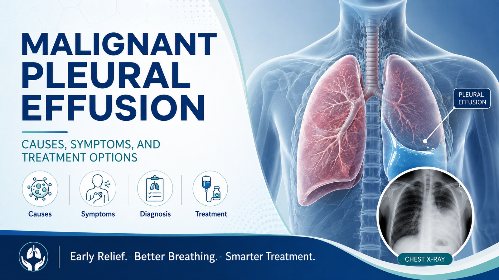 Malignant Pleural Effusion: Causes, Symptoms, and Treatment Options
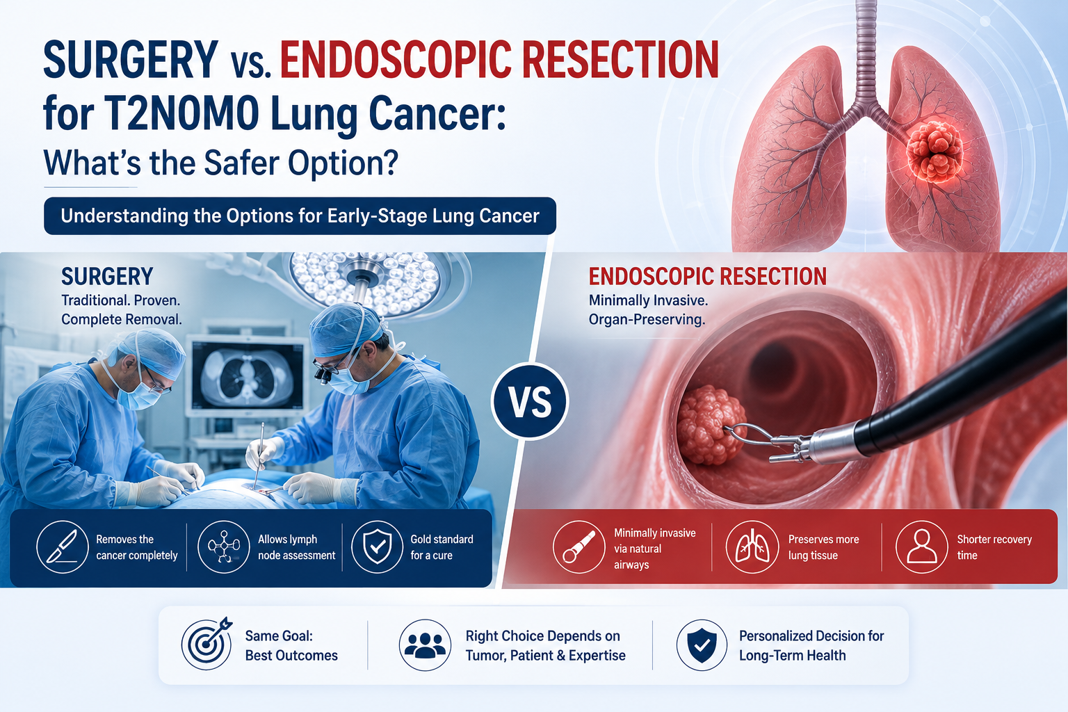 Surgery vs. Endoscopic Resection for T2N0M0 Lung Cancer: What’s the Safer Option?
 Robotic Lobectomy vs. Traditional VATS: An Expert’s Guide
Learn what causes malignant pleural effusion in lung cancer, its symptoms, and treatment options including thoracentesis, pleurodesis, and PleurX catheters.
T2N0M0 lung cancer diagnosis? Compare surgery vs. non-surgical options like SBRT. Learn about safety, survival rates, and what's the safer choice for you.
Robotic vs VATS lobectomy? An expert guide to choosing the best lung cancer treatment in Gurgaon & Delhi. Learn the pros, cons, and key differences.
Copyright 2026 © Dr .Parveen Yadav all rights reserved.
Proudly Scaled by Public Media Solution!