- Call : +91 9540210956
- Email: [email protected]
- Artemis Hospital Sector 51 Gurgaon
- 09Am - 5Pm


Fluid Around the Lung (Pleural Effusion): Causes and Treatment
- Home
- Blog Details

When a scan says there is fluid around the lung, many patients assume the lung itself is filling with water. That is not what a pleural effusion means. The fluid collects in the narrow space between the outside of the lung and the inside of the chest wall. This space normally contains only a small amount of fluid to allow the lung to slide as you breathe. When excess fluid builds up, the lung may not expand fully and breathlessness can follow.
A pleural effusion is a finding, not a final diagnosis. The next step is to understand why the fluid is there and whether it needs to be drained.
What Symptoms Can Fluid Around the Lung Cause?
A small effusion may cause no symptoms at all. Larger or quickly developing collections can lead to breathlessness, a dry cough, chest heaviness, fatigue, or pain that is worse on taking a deep breath. Some people notice that they are increasingly short of breath while walking, climbing stairs, or lying flat.
Symptoms such as fever, weight loss, night sweats, leg swelling, or coughing up blood may reflect the condition causing the fluid and are important to report. The amount of fluid on a scan does not always match how breathless a person feels, so the whole clinical picture matters.
Why Does Pleural Effusion Happen?
There are many causes. Pneumonia and other infections can inflame the pleura and cause fluid to collect. Heart failure, kidney disease, and liver disease can alter fluid balance in the body. Blood clots in the lung, autoimmune conditions, trauma, tuberculosis, and cancers can also cause an effusion.
Cancer is one possible cause, but it is not the only cause. No conclusion should be drawn from an X-ray alone. The right treatment depends entirely on the reason for the fluid.
How Is It Investigated?
A chest X-ray often detects pleural fluid. Ultrasound shows where the fluid lies and is commonly used to guide a needle safely. CT scanning gives a more detailed view of the lung, pleura, lymph nodes, and surrounding structures. Blood tests may provide additional clues.
When the cause is not already clear, a doctor may recommend pleural aspiration, also called thoracentesis. Local anaesthetic is used, and a thin needle or small catheter removes fluid for laboratory testing. This may also improve breathlessness. The sample can be examined for infection, inflammation, cancer cells, and other features that guide the next decision.
Why More Than One Test May Be Needed
Sometimes the first fluid sample gives a clear answer. Sometimes it does not. If concern remains after imaging and fluid analysis, the next step may be a repeat sample, image-guided pleural biopsy, or thoracoscopy. Thoracoscopy allows the specialist to look inside the pleural space and take tissue samples when needed.
This does not automatically mean a serious diagnosis. It means the team is trying to establish the cause accurately before recommending treatment.
Treatment Depends on the Cause
Small collections may be monitored if the underlying cause is known and being treated. Infection may require antibiotics, and infected fluid may need a chest drain. Fluid related to heart failure often improves with treatment aimed at removing excess body fluid. If an effusion is related to cancer or returns repeatedly, options can include repeat drainage, a longer-term pleural catheter, pleurodesis, or treatment directed at the cancer itself.
Drainage is not always the complete solution. If the cause is not treated, fluid may return. That is why the diagnostic part of the process is so important.
When to Seek Urgent Help
Sudden severe breathlessness, severe chest pain, fainting, confusion, blue lips, or coughing up more than a small amount of blood require urgent medical attention. Do not try to manage rapidly worsening breathlessness at home.
Questions to Ask Your Specialist
- How much fluid is present, and what is the likely cause?
- Do I need drainage now, and will it help my symptoms?
- Will the fluid be sent for testing?
- What follow-up imaging or appointments are planned?
- What should I do if breathlessness returns?
Final Thoughts
Pleural effusion is treatable, but the treatment must fit the cause. A clear assessment can relieve symptoms and prevent unnecessary delay in diagnosing infection, heart-related disease, or another underlying condition. Bring your scan images and reports to a chest specialist, not just the written result, so that the full picture can be reviewed.
What a Chest Drain Does
A chest drain is not required for every pleural effusion, but it is often used when there is a large amount of fluid, infected fluid, or a need for ongoing drainage. The tube sits in the pleural space and connects to a drainage system. The team monitors the amount and nature of the fluid, your breathing, and follow-up imaging before deciding when it can be removed.
When infection is suspected, drainage can be as important as antibiotics. Antibiotics circulate through the body, but thick infected fluid may not clear adequately without a way for it to leave the chest. This is one reason an effusion associated with pneumonia is assessed carefully rather than simply watched.
Follow-up Matters Even After Symptoms Improve
Breathlessness may improve soon after fluid is removed, but that does not by itself establish the cause. Keep the planned review and bring all fluid results and scan reports. If fluid returns, the timing and pattern of recurrence help the team decide whether another aspiration, a drain, pleurodesis, or more detailed testing is appropriate.
The aim is not only to make breathing easier today. It is to identify the reason for the fluid and choose a plan that is durable and appropriate for your overall health.

Dr. Parveen Yadav
18+ Yrs Exp | 5,700+ Thoracic & Robotic Cancer Surgeries
Dr. Parveen Yadav is a Director and Senior Consultant in Thoracic and Surgical Oncology, specializing in minimally invasive and robotic lung and esophageal surgeries, with advanced training from AIIMS and Tata Memorial Hospital.
View Full ProfileRecent Posts

Rib Pain When Coughing: Causes, Warning Signs and Treatment
- Dr. Parveen Yadav
- Oct 02, 2026

Food Stuck in Chest: Causes, Warning Signs and When to Get Help
- Dr. Parveen Yadav
- Sep 28, 2026
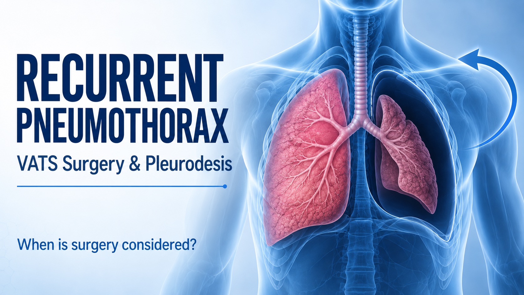
Recurrent Pneumothorax: When Are VATS and Pleurodesis Needed?
- Dr. Parveen Yadav
- Sep 24, 2026

Segmentectomy vs Lobectomy: Choosing Surgery for Early Lung Cancer
- Dr. Parveen Yadav
- Sep 20, 2026
Related Posts
Recurrent Pneumothorax: When Are VATS and Pleurodesis Needed?
Learn when recurrent pneumothorax needs surgery, how VATS and pleurodesis help prevent another collapsed lung, and what recovery and follow-up involve.
- Read More
- Sep, 24 2026

Preparing for Lung Surgery: A Patient Checklist
A practical, patient-first checklist for preparing for lung surgery, from tests and medicines to smoking cessation, support at home and questions for your surgeon.
- Read More
- Sep, 14 2026
Persistent Cough: When Should You See a Chest Specialist?
Most coughs settle on their own. Learn which symptoms, duration and risk factors mean a persistent cough should be evaluated by a chest specialist.
- Read More
- Sep, 11 2026
Copyright 2026 © Dr .Parveen Yadav all rights reserved.
Proudly Scaled by Public Media Solution!