


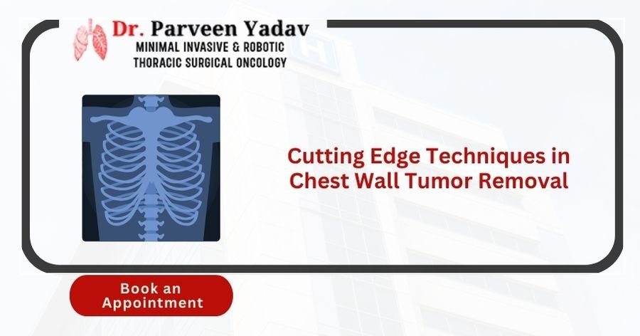
While chest wall tumours are relatively rare, they can be life-threatening and significantly impact a patient's overall well-being and quality of life. These tumours, which develop in the chest wall's bones, muscles, or soft tissues, can range from benign (non-cancerous) to malignant (cancerous). Thanks to advancements in medical technology, several cutting-edge techniques have emerged, offering less invasive procedures, quicker recovery times, and improved patient outcomes. This blog explains the latest chest wall tumour removal advancements and highlights how these innovative methods reshape patient care and recovery.
Understanding the different forms of chest wall tumours, whether primary or secondary, can empower patients with knowledge and a sense of control over their condition. Primary tumours can be further classified into benign ones like lipomas, fibromas, and neurofibromas and malignant ones like sarcomas, chondrosarcomas, and osteosarcomas. Secondary tumours often spread to the chest wall from cancers of the breast, lung, or kidney.
Common symptoms of chest wall tumours include localised pain, swelling, visible lumps, or discomfort while breathing. As these tumours grow, they can compress or invade surrounding tissues, affecting vital functions such as breathing and circulation.
Causes and risk factors for chest wall tumours may include genetic predisposition, prior radiation exposure, specific inherited syndromes, or trauma. While some risk factors are identifiable, the exact cause often remains unknown, making early diagnosis and intervention crucial.
Diagnosing a chest wall tumour involves multiple steps to accurately determine the tumour's nature, size, and extent. Advanced imaging methods play a crucial role in diagnosis, providing patients with a sense of relief and comfort:
X-rays and CT Scans: X-rays offer initial insight into the presence and size of the tumour, while CT scans provide detailed cross-sectional images of the chest.
Magnetic Resonance Imaging (MRI) offers a more detailed image, especially of soft tissue involvement, providing a clearer picture of tumour margins and their relationship with surrounding structures.
Positron Emission Tomography (PET) Scans: Helps assess the metabolic activity of the tumour, distinguishing between benign and malignant growths.
Biopsies: A biopsy involves taking a small tumour tissue sample to analyse under a microscope. This confirms whether the tumour is benign or malignant and determines the most effective treatment approach.
Molecular imaging and 3D Reconstruction: Advanced tools like molecular imaging and 3D reconstruction provide surgeons with a precise visualisation of the tumour, enabling tailored surgical planning.
These diagnostic tools have significantly evolved, enabling earlier detection and better treatment outcomes by offering detailed insights into the tumour's behaviour and spread. The future of chest wall tumour surgery is filled with hope and optimism, with advancements in AI, less invasive treatments, and precision medicine promising even better outcomes.
Traditional surgical techniques for chest wall tumour removal have primarily focused on open surgeries, where a large incision is made to access and remove the tumour:
Wide Local Excision: Remove the tumour and a margin of covering healthy tissue to confirm complete excision.
Partial Rib Resections: Removing part of the rib when the tumour has invaded bony structures.
Limitations of these traditional methods include longer recovery times, higher risk of complications such as infections, increased post-operative pain, and more significant cosmetic concerns due to large incisions.
Despite these challenges, traditional approaches are still highly relevant in cases involving significant or complex tumours or when tumours are located near critical structures where more extensive surgical exposure is needed.
With technological advancements, new techniques have emerged, providing more options for patients and improving surgical outcomes.
Minimally Invasive Surgery (MIS): This technique uses smaller incisions, leading to lowered trauma and faster recovery. Techniques like Video-Assisted Thoracoscopic Surgery (VATS) and Robotic-Assisted Surgery provide surgeons with enhanced precision. VATS, for example, uses a camera and specialised instruments inserted through small incisions, allowing for less invasive tumour removal.
Robotic Surgery for Chest Wall Tumors: Robotic surgery leverages advanced robotic systems that offer greater flexibility, precision, and control than traditional laparoscopic surgery. With robotic arms and 3D visualisation, surgeons can navigate complex anatomical structures more efficiently, reducing the risk of complications and improving patient outcomes. This method enhances skill, allowing for finer dissection and less damage to surrounding tissues.
3D-Printed Implants and Prosthetics: In cases where a significant portion of the chest wall must be removed, 3D-printed implants offer a personalised approach. Custom-made prosthetics are designed to fit the patient's unique anatomy, providing better structural support and improved cosmetic outcomes. These implants are made from biocompatible materials that integrate well with the body's natural tissues.
Cryoablation and Radiofrequency Ablation: These less invasive methods destroy cancer cells by using extreme cold (cryoablation) or heat (radiofrequency ablation). They are ideal for patients who cannot undergo surgery due to other health conditions. Cryoablation involves inserting a probe that freezes the tumour, while radiofrequency ablation uses heat generated by radio waves to target and destroy cancer cells.
Intraoperative Radiation Therapy (IORT): IORT is administered during surgery, targeting the tumour site directly with high doses of radiation. This approach minimises exposure to surrounding healthy tissues and helps reduce the risk of tumour recurrence. It is beneficial in cases where tumours are located near critical structures.
Advanced Imaging-Guided Surgery: Techniques like fluorescence imaging and augmented reality (AR) provide real-time guidance during surgery. Fluorescence imaging uses special dyes that bind to cancer cells, making them glow under specific lights. AR overlays virtual images on real-world views, helping surgeons see the exact tumour margins, enhancing precision, and reducing complications.
Reconstruction is critical to chest wall tumour surgery, as it helps restore function and appearance. Several innovative techniques are currently being utilised:
Biodegradable Meshes and Biological Implants: These materials temporarily support the chest wall, allowing natural tissue to regrow and strengthen the area over time.
Tissue Engineering: Involves cultivating new tissues in the lab to replace those lost during surgery, ensuring the chest wall remains structurally sound and functional.
Stem Cell Therapy: Stem cells can renew damaged tissues, providing a new frontier in post-surgical recovery. They help speed up healing, reduce scarring, and improve overall outcomes.
The adoption of these advanced techniques offers numerous benefits, including:
Improved Surgical Precision and Outcomes: Technologies like robotic surgery and advanced imaging allow for more precise tumour removal, reducing the risk of incomplete excision or damage to surrounding structures.
Reduced Recovery Time: Minimally invasive approaches, such as VATS and robotic surgery, result in faster hospital stays and speedier recovery times, enabling patients to resume their normal activities sooner.
Enhanced Quality of Life: Smaller incisions, reduced pain, and faster recovery improve patients' overall quality of life undergoing these procedures.
Lower Risk of Complications: These techniques minimise the risks associated with traditional open surgeries, such as infections, bleeding, and post-operative pain.
Selecting the most appropriate surgical technique involves careful consideration of various factors:
Tumor Type and Location: Depending on the type and location of the tumour, different techniques may be more suitable.
Patient Health: The patient's overall health status, age, and any existing medical conditions must be considered to choose the safest and most helpful approach.
Multidisciplinary Evaluation: A comprehensive assessment by a team of specialists, including oncologists, surgeons, and radiologists, ensures a personalised therapy program tailored to the patient's requirements.
Customised Treatment Plans: Customizing the treatment approach helps maximise outcomes and minimise complications, leading to a better recovery experience for the patient.
Post-surgical care is vital in ensuring a smooth recovery and reducing the risk of complications:
Physical Therapy and Rehabilitation are essential for restoring strength, mobility, and respiratory function. Rehabilitation schedules are tailored to individual needs to enhance recovery.
Pain Management: Effective pain control is crucial for patient comfort and recovery. A combination of medications, nerve blocks, and alternative therapies like acupuncture may be employed.
Regular Monitoring: Routine follow-up appointments help detect any signs of tumour recurrence or complications early, allowing for prompt intervention.
Nutritional Support and Lifestyle Adjustments: Proper nutrition supports healing and boosts immunity, while lifestyle modifications, such as quitting smoking, can improve overall health outcomes.
The future of chest wall tumour surgery is promising, with several emerging trends:
Artificial Intelligence (AI) and Machine Learning: AI can aid in surgical planning, predict outcomes, personalise treatment plans, and enhance precision and effectiveness.
Less Invasive Treatments: Research into new therapies like targeted drug delivery, immunotherapy, and gene therapy is ongoing, offering hope for less invasive and more effective treatments in the future.
Precision Medicine: Tailoring treatment plans based on genetic profiles and individual patient characteristics promises even better outcomes and reduced side effects.
The field of chest wall tumour surgery is rapidly advancing, offering patients a range of cutting-edge techniques that minimise invasiveness, reduce recovery times, and improve overall outcomes. Dr. Parveen Yadav and his team at Chest Surgery India are at the forefront of adopting these innovative methods, ensuring that every patient receives the most effective and personalised care possible.
1. How long does recovery take after chest wall tumour surgery?
Recovery time depends on the type of surgery, but minimally invasive techniques generally allow patients to recover within a few weeks.
2. What are the risks associated with chest wall tumour surgery?
Risks include infection, bleeding, and damage to nearby organs, but advanced techniques significantly minimise these risks.
3. How effective are minimally invasive surgeries for chest wall tumours?
Minimally invasive surgeries, such as VATS and robotic-assisted surgeries, are highly effective. They offer precise tumour removal with minimal damage to surrounding tissues.
4. Is robotic surgery right for all types of chest wall tumours?
Robotic surgery is ideal for many cases, but the suitability depends on the tumour's size, location, and complexity.
5. What are the options if a tumour recurs after initial surgery?
Treatment options may include additional surgery, radiation therapy, or targeted drug therapies, depending on the tumor's characteristics and the patient's health status.
Dr. Parveen Yadav is a highly recommended surgeon or specialist for chest wall tumour cancer in Gurgaon, Delhi. He specialises in minimally invasive and robotic thoracic onco surgery. He has been recognised for 18+ years as the best chest surgeon in India for his expertise in treating chest-related (Chest Surgery) ailments, such as Esophageal (Food Pipe Cancer), Lung, Tracheal (Throat), Chest wall tumours, Mediastinal Tumours, Empyema, and Bronchopleural Fistula cancer. With a focus on precision and innovation, he is dedicated to offering exceptional care to his patients, utilising techniques to ensure optimal outcomes.
18+ Yrs Exp | 5,700+ Thoracic & Robotic Cancer Surgeries
Dr. Parveen Yadav is a Director and Senior Consultant in Thoracic and Surgical Oncology, specializing in minimally invasive and robotic lung and esophageal surgeries, with advanced training from AIIMS and Tata Memorial Hospital.
View Full Profile Esophageal Cancer Surgery Recovery Timeline: First Week to 3 Months
 Second Opinion for Esophageal Cancer in India: When and Why It Matters
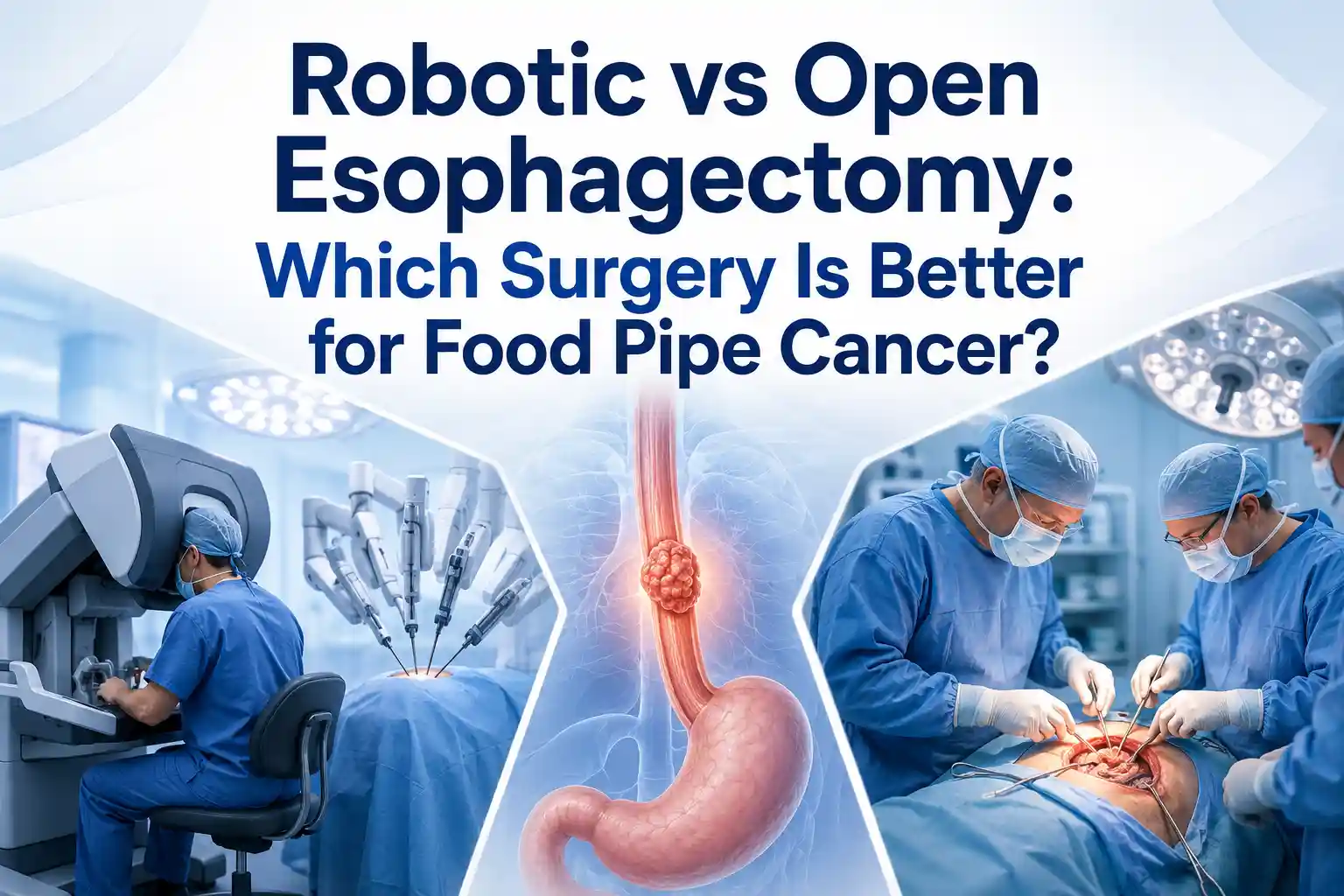 Robotic vs Open Esophagectomy: Which Surgery Is Better for Food Pipe Cancer?
Understand the esophageal cancer surgery recovery timeline from the first week to 3 months, including diet, pain, walking, breathing exercises, follow-up, and warning signs.
Learn when to take a second opinion for esophageal cancer in India, what reports to share, and why expert review matters before surgery, chemotherapy, or radiation.
Compare robotic vs open esophagectomy for food pipe cancer. Learn differences in pain, recovery, blood loss, lymph node clearance, hospital stay, cost, and when open surgery may be better.
Copyright 2026 © Dr .Parveen Yadav all rights reserved.
Proudly Scaled by Public Media Solution!