- Call : +91 9540210956
- Email: [email protected]
- Artemis Hospital Sector 51 Gurgaon
- 09Am - 5Pm


Does a Positive PET-CT Always Mean Lung Cancer?
- Home
- Blog Details
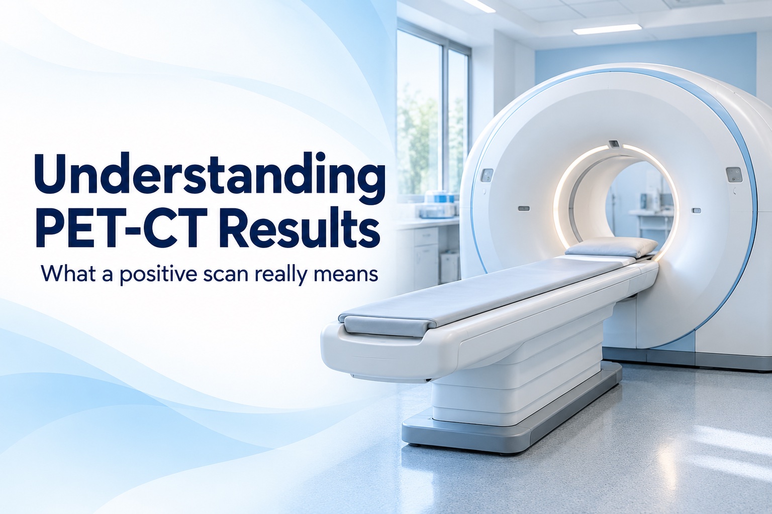
A PET-CT report that describes an “FDG-avid lung nodule” or “metabolically active lymph nodes” can sound like a confirmed cancer diagnosis. It is not that simple. PET-CT is an important investigation, but it measures tracer uptake rather than identifying cancer cells under a microscope.
A positive PET-CT can reflect lung cancer, infection, inflammation or changes related to treatment. The useful next question is not just whether an area lights up, but what that finding means alongside the CT appearance, previous scans, symptoms and any tissue results.
This guide explains the evidence behind false-positive and false-negative findings, why the Indian context matters, and how specialists decide between biopsy, surveillance and treatment. The references include professional guidelines and published research, with study limitations explained where numbers are used.
What PET and CT Each Contribute
The CT component maps structures: a nodule's location, size and relationship to the airways or chest wall. The PET component maps the distribution of a radioactive tracer. For many lung cancer investigations this is fluorodeoxyglucose, usually abbreviated to FDG, a substance similar to glucose.
Many cancer cells use glucose actively, so FDG uptake can make them more conspicuous. However, uptake is not unique to cancer. PET and CT complement one another: an area of increased uptake needs an anatomical explanation, and an abnormality on CT may remain relevant even when uptake is low. RadiologyInfo's PET-CT guide describes these different roles.
For a patient, “positive” is therefore a shorthand description, not a complete conclusion. Ask which exact finding the team considers suspicious and what would establish its cause.
Why Infection and Inflammation Can Light Up
Immune cells also consume glucose. When they gather in an infected or inflamed area, FDG uptake can increase. The EANM/SNMMI guideline on infection and inflammation explains this biological mechanism. In fact, PET is deliberately used to investigate some inflammatory and infectious diseases.
In chest imaging, possible explanations include tuberculosis, other infections and inflammatory conditions such as sarcoidosis. Healing tissues after surgery or radiation can also show uptake. The joint EANM/SNMMI/ESTRO recommendations for PET-CT in lung cancer discuss these sources of uptake unrelated to tumor.
This is why your team needs the dates of recent operations, biopsies, radiotherapy and significant infections. That history can change how a finding is interpreted. It does not, by itself, prove that a particular abnormality is benign.
Why TB and Other Infections Matter in India
In settings where infectious lung disease is common, a suspicious PET result can be harder to interpret. This is a practical reason to consider infection alongside cancer, rather than applying a result from a different population without context.
A 2014 JAMA meta-analysis by Deppen and colleagues included 70 studies and 8,511 lung nodules. The adjusted specificity was about 61% in populations with endemic infectious lung disease, compared with 77% in non-endemic populations. Specificity describes how often a test correctly classifies a non-cancerous finding as negative.
These figures do not mean that 39% of positive scans are false positives. The chance that a positive result represents cancer also depends on how common cancer is in the population being tested. The studies varied substantially, and older pooled results are not a personalised estimate for a modern scanner or an individual patient.
The clinical lesson is narrower: regional infection patterns affect interpretation. A history of TB should be shared, but neither TB nor cancer should be assumed from uptake alone.
Understanding the Words in Your Report
| Term | What it describes | Question to ask |
|---|---|---|
| FDG-avid | Tracer uptake in a tissue | What are the likely explanations here? |
| SUV or SUVmax | A numerical description of uptake, including the highest measured value | How does this fit the CT appearance? |
| Indeterminate | The finding cannot be confidently classified | Which test or follow-up will clarify it? |
| Suspicious for malignancy | The imaging raises concern about cancer | Is confirmation needed before treatment? |
Memorial Sloan Kettering's radiology terminology guide explains uptake measurements and uncertainty in reports. Read the report's overall impression and recommendations, rather than extracting one phrase from the findings section.
Why an SUV Cutoff Cannot Diagnose Cancer
You may see online advice that an SUV above 2.5 means cancer, or that a lower value means a benign nodule. That rule is unreliable. The British Thoracic Society pulmonary nodule guideline explicitly rejects the idea that an SUV below 2.5 reliably establishes a benign cause.
SUV is influenced by technical and biological conditions, including blood glucose, the interval between injection and imaging, and imaging methods. The EANM tumor-imaging procedure guideline explains why preparation and consistent acquisition matter for quantitative interpretation.
A practical example: comparing your SUV of 4 with a relative's SUV of 8 does not establish that your lesion is “half as serious.” These measurements do not form a cancer severity scale. Ask what the uptake adds to the assessment of your lesion, rather than trying to interpret the number independently.
Can a PET-Negative Finding Still Be Cancer?
Yes. PET has limits in detecting small lesions and tumors that take up relatively little FDG. A small abnormality can be difficult to separate from background activity. Certain tumors, including some carcinoid, mucinous and lepidic tumors, may show low uptake. Ground-glass nodules also need particular care in interpretation.
The BTS guideline and joint lung cancer PET recommendations discuss these limitations. A negative scan therefore has to be weighed against the CT findings and the pre-existing level of concern. It is not permission to abandon a planned nodule follow-up.
Our guide to a lung nodule found on CT explains the broader evaluation. Ask your specialist whether the lesion's size or appearance limits how much reassurance the PET result can provide.
How PET-CT and Biopsy Fit Together
PET-CT can help identify the extent of suspected disease and a useful place to sample. Biopsy examines cells or tissue and can provide information that imaging cannot. The appropriate route depends on the target: a lung lesion, an airway abnormality or a chest lymph node may need different approaches.
Cancer Research UK's lung cancer testing guide describes how imaging, bronchoscopy and tissue sampling contribute to the investigation. This is not a fixed sequence in which every patient needs every test.
For example, if a chest lymph node would change the proposed treatment, the team may focus on sampling that node rather than repeating a sample from an already diagnosed primary tumor. Ask what question the planned biopsy will answer and whether enough material is needed for further laboratory testing.
Do PET-Positive Lymph Nodes Prove Spread?
No. They may be suspicious, but inflammation and infection can affect nodes too. NICE guideline NG122 recommends systematic sampling of PET-positive or enlarged chest nodes with EBUS-TBNA, EUS-FNA or both when nodal status would affect treatment.
A negative sampling result is also interpreted in context. NICE advises considering surgical staging when suspicion remains high and the answer would change management. Our EBUS biopsy article explains the patient experience.
Similarly, if an isolated abnormality elsewhere would change a potentially curative plan, NICE recommends confirming it by biopsy or further imaging. The point is to establish a consequential finding before making a major treatment decision, not to biopsy every area of uptake.
What Could Happen After the Results Review?
The consultation should leave you with a specific plan. Depending on the combined findings, that might involve tissue sampling, additional imaging, comparison with older scans, or a scheduled surveillance pathway. If a diagnosis has been established, the next discussion may concern treatment and fitness assessment.
Ask the team to distinguish three things: what is known, what is suspected, and what remains unresolved. “Review later” should come with an interval, a responsible clinician and a way to receive the result. Write these down so uncertainty does not turn into an unplanned delay.
A second opinion is most useful when the reviewer can see the original images and pathology, not just a screenshot of the PET conclusion.
Preparing for a PET-CT or a Results Appointment
For the scan itself, follow the department's fasting and activity instructions and arrange a specific plan if you have diabetes. Do not alter insulin or other medicines on your own. Blood glucose and recent exertion can affect tracer distribution, which is one reason standardised preparation matters.
NHS information on PET scanning covers preparation and the need to tell the department about pregnancy or breastfeeding. The department should also provide instructions for the period after the scan.
For the results consultation, bring:
- The PET-CT images and full written report.
- Previous CT or PET-CT images with dates.
- Biopsy reports and any available laboratory results.
- A timeline of recent infection, surgery, biopsy and cancer treatment.
- A medicine list and your most important questions.
Frequently Asked Questions
Can PET-CT distinguish TB from lung cancer?
Not reliably in every case. Both can show uptake. Ask whether the proposed sampling and laboratory tests will investigate the relevant possibilities rather than assuming one explanation.
Is a higher SUV always a more advanced cancer?
No. Uptake intensity is not the same as cancer stage. Ask your doctor to explain the extent of disease separately from the uptake measurement.
Does every positive scan need immediate surgery?
No. The next step depends on what remains uncertain and which result would change treatment. Ask why the recommended investigation is the best next step for you.
Can I cancel follow-up if I feel well?
No. Feeling well does not resolve a scan finding. Keep the planned review and ask for a clear explanation of any surveillance schedule.
Getting a Thoracic Surgical Opinion
A useful review should connect your scan to a decision: whether confirmation is needed, which target to sample, and what the possible results would mean. If you need a thoracic surgical opinion, book a consultation with Dr. Parveen Yadav and bring the complete investigation record.
Selected Research and Guidelines
- Deppen et al., JAMA, 2014: meta-analysis of PET accuracy in populations with infectious lung disease; useful context, not an individual probability calculator.
- BTS pulmonary nodule guideline, 2015: limitations of PET, small nodules and fixed SUV thresholds.
- Joint EANM/SNMMI/ESTRO lung cancer PET recommendations, 2022: tumor and non-tumor causes of uptake.
- NICE NG122: confirmation of findings that influence staging and treatment.

Dr. Parveen Yadav
18+ Yrs Exp | 5,700+ Thoracic & Robotic Cancer Surgeries
Dr. Parveen Yadav is a Director and Senior Consultant in Thoracic and Surgical Oncology, specializing in minimally invasive and robotic lung and esophageal surgeries, with advanced training from AIIMS and Tata Memorial Hospital.
View Full ProfileRecent Posts
Lung Function Tests Before Lung Surgery: FEV1, DLCO and Fitness Explained
- Dr. Parveen Yadav
- Oct 17, 2026

Chest Drain After Lung Surgery: Care, Removal and Recovery
- Dr. Parveen Yadav
- Oct 10, 2026
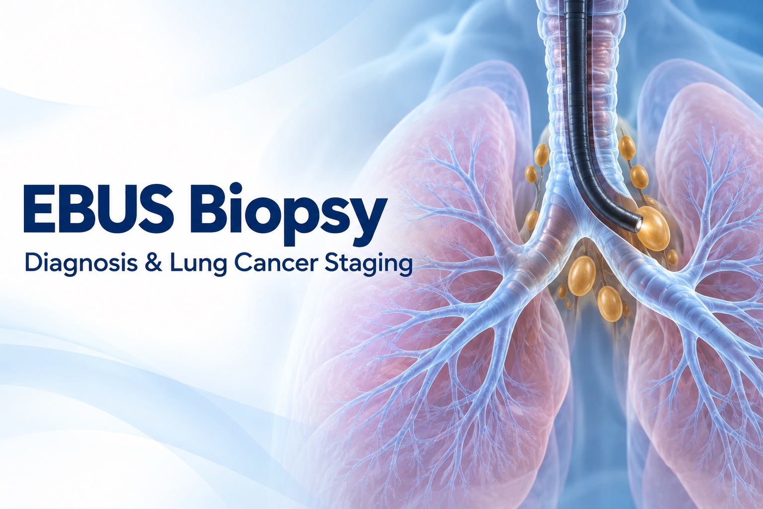
EBUS Biopsy: What to Expect and How It Helps Stage Lung Cancer
- Dr. Parveen Yadav
- Oct 06, 2026
Rib Pain When Coughing: Causes, Warning Signs and Treatment
- Dr. Parveen Yadav
- Oct 02, 2026
Related Posts
EBUS Biopsy: What to Expect and How It Helps Stage Lung Cancer
Learn why EBUS biopsy is used to sample chest lymph nodes, how to prepare, what recovery involves and how results help guide lung cancer treatment.
- Read More
- Oct, 06 2026
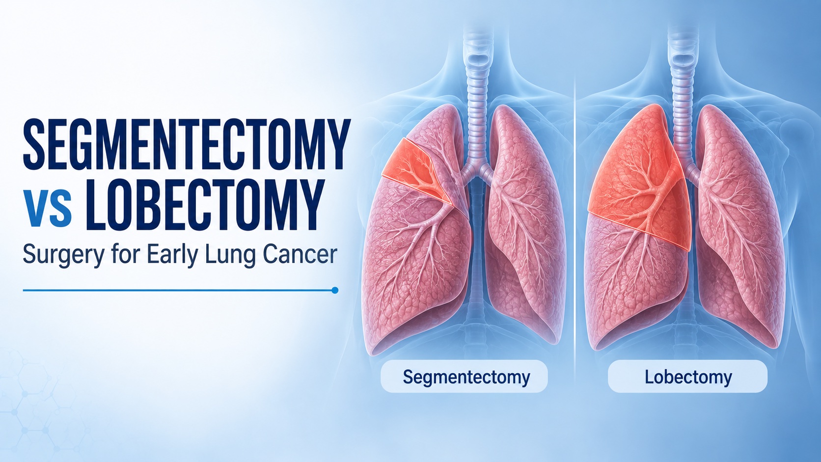
Segmentectomy vs Lobectomy: Choosing Surgery for Early Lung Cancer
Compare segmentectomy and lobectomy for early lung cancer: who may suit lung-sparing surgery, why lymph nodes matter, and what to ask your thoracic surgeon.
- Read More
- Sep, 20 2026
A Lung Nodule on CT Scan: What Happens Next?
A clear guide to lung nodules found on CT scans: what they are, why many are benign, and how specialists decide on follow-up, PET-CT or biopsy.
- Read More
- Sep, 08 2026
Copyright 2026 © Dr .Parveen Yadav all rights reserved.
Proudly Scaled by Public Media Solution!