


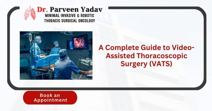
Advancements in surgical procedures have made it possible to offer less invasive options to patients for treating thoracic conditions. Video-assisted thoracoscopic surgery (VATS) is one modern technique that has revolutionised chest surgeries.
Traditional open chest surgeries usually require large incisions, causing more pain and more extended recovery periods. However, VATS, a minimally invasive procedure, permits surgeons to perform complicated surgeries through small incisions with less postoperative pain and quicker recovery times.
Advancements in advanced medical technology have led to the international use of VATS for diagnosing and treating different chest conditions. This blog will cover all aspects of VATS, including the procedure, its benefits, limitations, and future developments.
Whether you're a patient considering this surgery, this article will help you understand everything you need about VATS and why it is becoming the preferred method for thoracic procedures.
Video-assisted thoracoscopic surgery (VATS) is a minimally invasive process used to diagnose and treat chest diseases such as lung cancer, pleural effusion, and pneumothorax. Unlike traditional open chest surgeries, which involve making a large incision and spreading the ribs, VATS uses a thin thoracoscope inserted through a few small incisions (typically 2-4 cm in size). The thoracoscope has a small video camera that displays a high-definition image of the surgical area on a monitor, letting the surgeon guide and operate precisely.
Smaller Incisions: Typically 3 to 4 small incisions.
Thoracoscope: A camera-equipped tube that offers a clear view of the chest cavity.
Minimally Invasive Tools: Remove tissue, biopsy, or perform repairs.
Real-Time Visual Guidance: Surgeons use a monitor to view and manipulate tissues.
This approach causes less trauma to the muscles and tissues surrounding the ribs, resulting in reduced postoperative pain, more minor scars, and quicker recovery compared to traditional thoracotomy (open chest surgery).
Traditional thoracic surgeries, also known as thoracotomies, are more invasive, requiring a big incision along the side of the chest and the spreading of ribs to provide access to the lungs, esophagus, or other chest organs. This procedure often results in intense postoperative pain and a more extended recovery period due to the trauma caused to muscles, tissues, and ribs.
On the other hand, VATS is performed using several small incisions (ports), through which a thoracoscope and surgical instruments are inserted. This allows the surgeon to execute the same procedures with a much smaller incision and less trauma to the surrounding areas. Additionally, VATS can be performed with the patient under general anaesthesia, provided they are comfortable and pain-free during the procedure.
|
Feature |
Traditional Thoracic Surgery |
Video-Assisted Thoracoscopic Surgery (VATS) |
|
Incision Size |
Large incision (10-20 cm) |
Small incisions (2-4 cm) |
|
Rib Spreading |
Requires rib spreading, causing more trauma |
No rib spreading, minimal tissue damage |
|
Postoperative Pain |
High levels of pain due to muscle and rib involvement |
Reduced pain due to smaller incisions |
|
Scarring |
Large, visible scar |
Small, less noticeable scars |
|
Hospital Stay |
5 to 10 days or longer |
2 to 5 days |
|
Recovery Time |
6 to 8 weeks or more |
2 to 4 weeks |
|
Risk of Complications |
Higher risk of infection, bleeding, and lung problems |
Lower risk due to minimally invasive nature |
|
Patient Suitability |
Suitable for complex and large tumours |
Suitable for early-stage cancers and localized conditions |
|
Surgical Visibility |
Direct visibility of organs |
Indirect visibility using a camera |
|
Surgeon Expertise |
Requires standard surgical skills |
Requires specialized training and expertise in VATS |
|
Overall Outcome |
Effective but with more trauma and longer recovery |
Effective with reduced trauma and quicker recovery |
"minimally invasive" describes any procedure that minimizes surgical trauma to the patient. VATS is considered minimally invasive because it utilises small incisions and specialised instruments, leading to:
Reduced Postoperative Pain: Smaller cuts mean less tissue damage.
Lower Risk of Complications: Minimal incisions lessen the chance of infections and other complications.
Faster Recovery Time: Patients can continue normal activities sooner.
Improved Cosmetic Results: Smaller incisions indicate more minor scars.
These advantages make VATS a preferred choice for patients and surgeons, especially for those who wish to avoid the long-term impacts of traditional open surgeries.
VATS has become the go-to option for various thoracic conditions, both benign and malignant. Some of the primary conditions treated include:
Lung Cancer: VATS is used for both early-stage lung cancer (lobectomy) and diagnostic purposes (biopsy).
Pleural Effusion: VATS can drain excess fluid from the pleural space.
Pneumothorax (Collapsed Lung): VATS effectively treats persistent or recurrent pneumothorax by repairing the damaged areas of the lung.
Empyema: Used to remove infected fluid and pus from the pleural cavity.
Mediastinal Tumors: Removal of tumours in the mediastinum (the area between the lungs).
Oesophagal Conditions: Some oesophagal surgeries can also be performed using VATS, reducing complications.
A complete preoperative evaluation confirms the patient's fitness for surgery. Tests include:
Chest X-rays and CT Scans: To determine the location and size of the condition.
Blood Tests: To consider the patient's overall health.
Pulmonary Function Tests: To asses lung capacity and function.
The VATS procedure follows a systematic approach:
Anesthesia Administration: General anaesthesia is given to keep the patient unconscious and pain-free.
Port Placement: Small incisions are made, and ports are inserted.
Thoracoscope Insertion: The thoracoscope is inserted through one port, allowing the surgeon to visualize the inside of the chest cavity.
Surgical Manipulation: The surgeon performs the necessary process, such as removing a part of the lung, draining fluid, or taking a biopsy using specialised instruments.
Closure: After the procedure, the instruments are removed, and the incisions are sealed using sutures.
Patients are moved to the recovery room, where vital signs are monitored closely. Pain management, respiratory exercises, and early mobilization are the focus to speed up recovery.
1. Less Pain: Minimizes muscle and tissue trauma.
2. Quicker Recovery: Patients can often return to work and regular activities within a few weeks.
3. Better Outcomes for Lung Cancer: Examinations show comparable or even better outcomes for early-stage lung cancer patients.
4. Cosmetic Benefits: The small scars are much less noticeable.
1. Learning Curve: Requires specialized training and experience for surgeons.
2. Limited Visibility: This can be challenging for complex cases.
3. Risk of Conversion to Open Surgery: If complications arise, the surgeon may sometimes need to switch to traditional surgery.
VATS is ideal for patients with:
Early-stage lung cancer
Pleural diseases
Localized tumours in the chest cavity
However, patients with extensive adhesions, previous thoracic surgeries, or poor lung function may not be appropriate for VATS and might require traditional thoracotomy.
While VATS is generally safe, it does carry some risks, such as:
Bleeding: From blood vessels or organs.
Air Leaks: Persistent air leaks from the lung tissue.
Infection: At the incision site or inside the chest.
However, the overall complication risk is significantly lower than traditional thoracic surgeries.
Depending on the complexity or difficulty of the surgery, patients can typically continue light activities in 2 weeks and fully recover within 4 to 6 weeks.
Pain Medication: Pain is managed with oral and intravenous medications.
Breathing Exercises: Expanding the lungs and preventing complications like pneumonia is essential.
Patients are motivated to consume a diet rich in proteins, fruits, and vegetables to facilitate healing and avoid strenuous activities until fully recovered.
The success rate of VATS is high, particularly for early-stage lung cancers and benign conditions. Studies show that VATS patients have a 5-year survival rate similar to open surgeries, with rarer complications and improved quality of life.
Video-assisted thoracoscopic Surgery (VATS) is a revolutionary technique offering significant advantages over traditional open chest surgeries. Its minimally invasive nature, shorter recovery times, and smaller incisions have made it a favoured option for patients and surgeons. However, successful outcomes depend on the surgeon's experience and patient selection. If you're looking for VATS, Dr. Parveen Yadav at Chest Surgery India is a leading expert in performing VATS procedures with exceptional results.
1. Is Video-Assisted Thoracoscopic Surgery (VATS) safe?
Yes, VATS is considered safe and has fewer complications compared to traditional thoracic surgeries.
2. How long does it take to recover from VATS?
Most patients fully recover within 4 to 6 weeks, depending on the procedure.
3. What is the success rate of VATS?
VATS has a high success rate, particularly for treating early-stage lung cancer.
4. What are the risks associated with VATS?
Risks include bleeding, infection, and minor lung injuries, but these are rare.
5. How does VATS differ from robotic surgery?
While both are minimally invasive, robotic surgery offers greater precision with robotic arms, making it suitable for more complex cases.
Dr. Parveen Yadav is a highly recommended surgeon or specialist for Video-assisted thoracoscopic surgery (VATS) in Gurgaon, Delhi. He specialises in minimally invasive and robotic thoracic onco surgery. He has been recognised for 18+ years as the best chest surgeon in India for his expertise in treating chest-related (Chest Surgery) ailments, such as Esophageal (Food Pipe Cancer), Lung, Tracheal (Throat), Chest wall tumours, Mediastinal Tumours, Empyema, and Bronchopleural Fistula cancer. With a focus on precision and innovation, he is dedicated to offering exceptional care to his patients, utilising techniques to ensure optimal outcomes.
18+ Yrs Exp | 5,700+ Thoracic & Robotic Cancer Surgeries
Dr. Parveen Yadav is a Director and Senior Consultant in Thoracic and Surgical Oncology, specializing in minimally invasive and robotic lung and esophageal surgeries, with advanced training from AIIMS and Tata Memorial Hospital.
View Full Profile Esophageal Cancer Surgery Recovery Timeline: First Week to 3 Months
 Second Opinion for Esophageal Cancer in India: When and Why It Matters
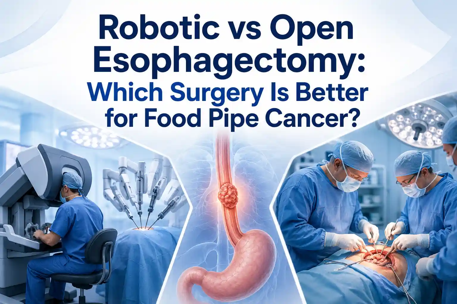 Robotic vs Open Esophagectomy: Which Surgery Is Better for Food Pipe Cancer?
Understand the esophageal cancer surgery recovery timeline from the first week to 3 months, including diet, pain, walking, breathing exercises, follow-up, and warning signs.
Learn when to take a second opinion for esophageal cancer in India, what reports to share, and why expert review matters before surgery, chemotherapy, or radiation.
Compare robotic vs open esophagectomy for food pipe cancer. Learn differences in pain, recovery, blood loss, lymph node clearance, hospital stay, cost, and when open surgery may be better.
Copyright 2026 © Dr .Parveen Yadav all rights reserved.
Proudly Scaled by Public Media Solution!