


Esophageal cancer requires distinct approaches for diagnosis and treatment due to its various types. This comprehensive guide will delve into the primary kinds of esophageal cancer, focusing on the critical differences between Adenocarcinoma and Squamous Cell Carcinoma. Additionally, we'll explore rare types and variants, shedding light on the less common but equally important aspects of this malignancy. Also, look at our complete blog on Esophageal Cancer: Causes, Symptoms, Diagnosis, and Treatment.
In the Western world, Adenocarcinoma is the most common form. It typically originates in the glandular cells of the lower esophagus and is closely associated with gastroesophageal reflux disease (GERD). Risk factors include obesity, smoking, and Barrett's esophagus. Early symptoms may be subtle, making regular screenings crucial for early detection.
Squamous cell carcinoma, on the other hand, arises from the flat, thin cells lining the esophagus. This type is more common in Eastern countries and is often linked to factors such as smoking and excessive alcohol consumption. Symptoms may include difficulty swallowing, weight loss, and chest pain. Prevention strategies involve lifestyle changes and minimizing risk factors.
While Adenocarcinoma and Squamous Cell Carcinoma represent the majority of esophageal cancer cases, several rare types and variants exist, demanding specialized attention:
This aggressive variant is less common but tends to grow and spread rapidly. Its treatment approach may differ from more common types of esophageal cancer.
Adenosquamous carcinoma presents unique challenges in diagnosis and treatment planning by combining Adenocarcinoma and squamous cell carcinoma features.
Originating from smooth muscle cells, leiomyosarcoma is a rare form of esophageal cancer that requires a distinct therapeutic approach.
While generally rare, neuroendocrine tumors can affect the esophagus. These tumours arise from cells that produce hormones and may have a different prognosis than other types.
1. What are the common symptoms?
Ans: Esophageal cancer symptoms may include difficulty swallowing, unintentional weight loss, chest pain, and persistent coughing.
2. How is it diagnosed?
Ans: Diagnosis involves endoscopy, imaging tests, and biopsy to confirm the presence of cancerous cells.
3. Could you please clarify the risk factors that increase the chances of developing esophageal cancer?
Ans: Risk factors include smoking, excessive alcohol consumption, obesity, GERD, and specific dietary factors.
4. How is the stage determined?
Ans: Staging assesses tumour size, invasion extent, and metastasis.
5. What treatment options are available?
Ans: Treatment options include surgery, chemotherapy, radiation therapy, and targeted therapy, depending on the type and stage of cancer.
Finally, early detection and effective treatment of esophageal cancer rely on a comprehensive understanding of its nuances. If you or a loved one experiences symptoms or possesses risk factors, seeking medical advice promptly is essential. Dr. Parveen Yadav and Chest Surgery India are pillars of expertise and compassion that provide advanced patient care. Stay informed, stay vigilant, and together, we can conquer the challenges posed by this formidable disease.
18+ Yrs Exp | 5,700+ Thoracic & Robotic Cancer Surgeries
Dr. Parveen Yadav is a Director and Senior Consultant in Thoracic and Surgical Oncology, specializing in minimally invasive and robotic lung and esophageal surgeries, with advanced training from AIIMS and Tata Memorial Hospital.
View Full Profile Esophageal Cancer Surgery Recovery Timeline: First Week to 3 Months
 Second Opinion for Esophageal Cancer in India: When and Why It Matters
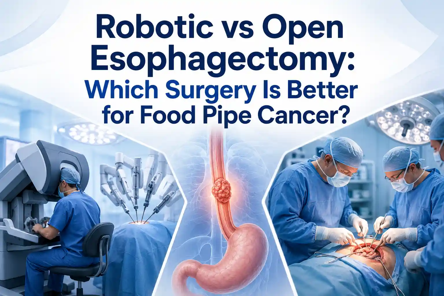 Robotic vs Open Esophagectomy: Which Surgery Is Better for Food Pipe Cancer?
Understand the esophageal cancer surgery recovery timeline from the first week to 3 months, including diet, pain, walking, breathing exercises, follow-up, and warning signs.
Learn when to take a second opinion for esophageal cancer in India, what reports to share, and why expert review matters before surgery, chemotherapy, or radiation.
Compare robotic vs open esophagectomy for food pipe cancer. Learn differences in pain, recovery, blood loss, lymph node clearance, hospital stay, cost, and when open surgery may be better.
Copyright 2026 © Dr .Parveen Yadav all rights reserved.
Proudly Scaled by Public Media Solution!